Mass: 18.015 Da / Num. of mol.: 1289 / Source method: isolated from a natural source / Formula: H2O
Sequence details
AUTHORS STATE THAT THE CORRECT SEQUENCE HAS A GLN AT POSITION 126. THE CORRECT SEQUENCE HAS BEEN ...AUTHORS STATE THAT THE CORRECT SEQUENCE HAS A GLN AT POSITION 126. THE CORRECT SEQUENCE HAS BEEN DEPOSITED TO THE GENBANK.
-
Experimental details
-
Experiment
Experiment
Method: X-RAY DIFFRACTION / Number of used crystals: 1
-
Sample preparation
Crystal
Density Matthews: 2.51 Å3/Da / Density % sol: 50.99 %
Crystal grow
Temperature: 298 K / Method: vapor diffusion, hanging drop / pH: 7.4 Details: 35% PEG 4K, 5% saturated Urea, 200 mM immidazole malate , pH 7.4, VAPOR DIFFUSION, HANGING DROP, temperature 298K
-
Data collection
Diffraction
Mean temperature: 100 K
Diffraction source
Source: SYNCHROTRON / Site: ALS / Beamline: 5.0.3 / Wavelength: 0.979764 Å
 Movie
Movie Controller

 Yorodumi
 Open data
 Basic information
 Components
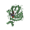
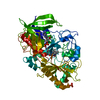
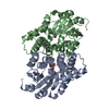
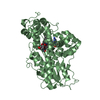
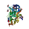
 Photolyase
  Keywords
 Function and homology information
 Authors
 Citation
 Structure visualization
 Downloads & links
 Other downloads


 PDBj
PDBj
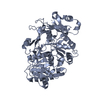
 Assembly
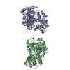


 Mass: 62.068 Da / Num. of mol.: 6 / Source method: obtained synthetically / Formula: C2H6O2
 Mass: 785.550 Da / Num. of mol.: 2 / Source method: obtained synthetically / Formula: C27H33N9O15P2 / Comment: FAD*YM
 Mass: 60.055 Da / Num. of mol.: 9 / Source method: obtained synthetically / Formula: CH4N2O
 Mass: 18.015 Da / Num. of mol.: 1289 / Source method: isolated from a natural source / Formula: H2O
 Sample preparation
 / Beamline: 5.0.3 / Wavelength: 0.979764 Å
 Processing