 Movie
Movie Controller

[English] 日本語
 Yorodumi
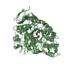
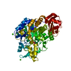
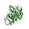
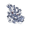
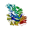
- PDB-1aq6: STRUCTURE OF L-2-HALOACID DEHALOGENASE FROM XANTHOBACTER AUTOTROPHICUS -
+ Open data

- Basic information
| Entry | Database: PDB / ID: 1aq6 | ||||||
|---|---|---|---|---|---|---|---|
| Title | STRUCTURE OF L-2-HALOACID DEHALOGENASE FROM XANTHOBACTER AUTOTROPHICUS | ||||||
 Components Components | L-2-HALOACID DEHALOGENASE | ||||||
 Keywords Keywords |  DEHALOGENASE / L-2-HALOACID DEHALOGENASE DEHALOGENASE / L-2-HALOACID DEHALOGENASE | ||||||
| Function / homology |  Function and homology information Function and homology information | ||||||
| Biological species |  Xanthobacter autotrophicus (bacteria) Xanthobacter autotrophicus (bacteria) | ||||||
| Method | X-RAY DIFFRACTION / SYNCHROTRON / MIR / Resolution: 1.95 Å | ||||||
 Authors Authors | Ridder, I.S. / Rozeboom, H.J. / Kalk, K.H. / Dijkstra, B.W. | ||||||
 Citation Citation | Journal: J.Biol.Chem. / Year: 1997 Title: Three-dimensional structure of L-2-haloacid dehalogenase from Xanthobacter autotrophicus GJ10 complexed with the substrate-analogue formate. Authors: Ridder, I.S. / Rozeboom, H.J. / Kalk, K.H. / Janssen, D.B. / Dijkstra, B.W. #1: Journal: Protein Sci. / Year: 1995Title: Crystallization and Preliminary X-Ray Analysis of L-2-Haloacid Dehalogenase from Xanthobacter Autotrophicus Gj10 Authors: Ridder, I.S. / Rozeboom, H.J. / Kingma, J. / Janssen, D.B. / Dijkstra, B.W. | ||||||
| History |
|
- Structure visualization
| Structure viewer | Molecule: MolmilJmol/JSmol |
|---|
- Downloads & links
-Download
| PDBx/mmCIF format | 1aq6.cif.gz | 111 KB | Display | PDBx/mmCIF format |
|---|---|---|---|---|
| PDB format | pdb1aq6.ent.gz | 85.9 KB | Display | PDB format |
| PDBx/mmJSON format | 1aq6.json.gz | Tree view | PDBx/mmJSON format | |
| Others |  Other downloads Other downloads |
-Validation report
| Arichive directory | https://data.pdbj.org/pub/pdb/validation_reports/aq/1aq6ftp://data.pdbj.org/pub/pdb/validation_reports/aq/1aq6 | HTTPS FTP |
|---|
-Related structure data
| Similar structure data |
|---|


-Links
 PDBj
PDBj

- Assembly
| Deposited unit | 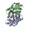
| ||||||||
|---|---|---|---|---|---|---|---|---|---|
| 1 |
| ||||||||
| Unit cell |
| ||||||||
| Noncrystallographic symmetry (NCS) | NCS oper: (Code: given Matrix: (-0.996674, -0.007367, 0.08116), Vector : |
-Components
| #1: Protein | Mass: 27494.373 Da / Num. of mol.: 2 / Source method: isolated from a natural source Details: SUBSTRATE ANALOGUE FORMATE PRESENT IN BOTH ACTIVE SITES Source: (natural) Xanthobacter autotrophicus (bacteria) / Strain: GJ10 / References: UniProt: Q60099, (S)-2-haloacid dehalogenase#2: Chemical | Formic acid  Mass: 46.025 Da / Num. of mol.: 3 / Source method: obtained synthetically / Formula: CH2O2 Mass: 46.025 Da / Num. of mol.: 3 / Source method: obtained synthetically / Formula: CH2O2#3: Water | ChemComp-HOH / | Water Mass: 18.015 Da / Num. of mol.: 334 / Source method: isolated from a natural source / Formula: H2O Mass: 18.015 Da / Num. of mol.: 334 / Source method: isolated from a natural source / Formula: H2O |
|---|
-Experimental details
-Experiment
| Experiment | Method: X-RAY DIFFRACTION / Number of used crystals: 1 |
|---|
- Sample preparation
| Crystal | Density Matthews: 2 Å3/Da / Density % sol: 38 % | ||||||||||||||||||||||||||||||||||||||||
|---|---|---|---|---|---|---|---|---|---|---|---|---|---|---|---|---|---|---|---|---|---|---|---|---|---|---|---|---|---|---|---|---|---|---|---|---|---|---|---|---|---|
| Crystal grow | Method: macroseeding / pH: 7 Details: MACROSEEDING, DROP: 16% PEG8000, 200 MM SODIUM FORMATE, 100 MM BIS-TRIS PH 7.0; WELL: 22% PEG8000, 200 MM SODIUM FORMATE, 100 MM BIS-TRIS PH 7.0, macroseeding | ||||||||||||||||||||||||||||||||||||||||
| Crystal grow | *PLUS Method: vapor diffusion, hanging drop / Details: used to seeding / PH range low: 7 / PH range high: 6.8 | ||||||||||||||||||||||||||||||||||||||||
| Components of the solutions | *PLUS
|
-Data collection
| Diffraction | Mean temperature: 120 K |
|---|---|
| Diffraction source | Source: SYNCHROTRON / Site: EMBL/DESY, HAMBURG  / Beamline: BW7B / Wavelength: 0.883 / Beamline: BW7B / Wavelength: 0.883 |
| Detector | Type: MARRESEARCH / Detector: IMAGE PLATE / Date: Jul 30, 1996 |
| Radiation | Monochromatic (M) / Laue (L): M / Scattering type: x-ray |
| Radiation wavelength | Wavelength: 0.883 Å / Relative weight: 1 |
| Reflection | Resolution: 1.95→25 Å / Num. obs: 32507 / % possible obs: 99.4 % / Observed criterion σ(I): 3 / Redundancy: 13.5 % / Rmerge(I) obs: 0.042 / Net I/σ(I): 15.2 |
| Reflection shell | Resolution: 1.95→1.98 Å / Redundancy: 13.5 % / Rmerge(I) obs: 0.156 / Mean I/σ(I) obs: 6 / % possible all: 98.7 |
| Reflection | *PLUS Num. measured all: 438845 |
- Processing
| Software |
| ||||||||||||||||||||||||||||||||||||||||||||||||||||||||||||||||||||||||||||||||||||
|---|---|---|---|---|---|---|---|---|---|---|---|---|---|---|---|---|---|---|---|---|---|---|---|---|---|---|---|---|---|---|---|---|---|---|---|---|---|---|---|---|---|---|---|---|---|---|---|---|---|---|---|---|---|---|---|---|---|---|---|---|---|---|---|---|---|---|---|---|---|---|---|---|---|---|---|---|---|---|---|---|---|---|---|---|---|
| Refinement | Method to determine structure: MIR / Resolution: 1.95→5 Å / Cross valid method: THROUGHOUT / σ(F): 0
| ||||||||||||||||||||||||||||||||||||||||||||||||||||||||||||||||||||||||||||||||||||
| Displacement parameters | Biso mean: 21.6 Å2 | ||||||||||||||||||||||||||||||||||||||||||||||||||||||||||||||||||||||||||||||||||||
| Refine analyze | Luzzati coordinate error obs: 0.2 Å / Luzzati d res low obs: 5 Å | ||||||||||||||||||||||||||||||||||||||||||||||||||||||||||||||||||||||||||||||||||||
| Refinement step | Cycle: LAST / Resolution: 1.95→5 Å
| ||||||||||||||||||||||||||||||||||||||||||||||||||||||||||||||||||||||||||||||||||||
| Refine LS restraints |
| ||||||||||||||||||||||||||||||||||||||||||||||||||||||||||||||||||||||||||||||||||||
| Software | *PLUS Name: REFMAC / Classification: refinement | ||||||||||||||||||||||||||||||||||||||||||||||||||||||||||||||||||||||||||||||||||||
| Refinement | *PLUS Rfactor Rwork: 0.19 | ||||||||||||||||||||||||||||||||||||||||||||||||||||||||||||||||||||||||||||||||||||
| Solvent computation | *PLUS | ||||||||||||||||||||||||||||||||||||||||||||||||||||||||||||||||||||||||||||||||||||
| Displacement parameters | *PLUS |
