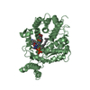
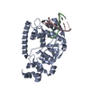
Entry Database : PDB / ID : 5c5hTitle R195K E. coli MenE with bound OSB-AMS 2-succinylbenzoate--CoA ligase Keywords Function / homology Biological species Escherichia coli (E. coli)Method / / / Resolution : 2.401 Å Authors Matarlo, J.S. / Shek, R. / Rajashankar, K.R. / Tonge, P.J. / French, J.B. Funding support Organization Grant number Country National Institutes of Health/National Institute of General Medical Sciences (NIH/NIGMS) GM102864 National Institutes of Health/National Institute of General Medical Sciences (NIH/NIGMS) GM103403
Journal : Biochemistry / Year : 2015Title : Mechanism of MenE Inhibition by Acyl-Adenylate Analogues and Discovery of Novel Antibacterial Agents.Authors : Matarlo, J.S. / Evans, C.E. / Sharma, I. / Lavaud, L.J. / Ngo, S.C. / Shek, R. / Rajashankar, K.R. / French, J.B. / Tan, D.S. / Tonge, P.J. History Deposition Jun 19, 2015 Deposition site / Processing site Revision 1.0 Oct 7, 2015 Provider / Type Revision 1.1 Nov 4, 2015 Group Revision 1.2 Sep 6, 2017 Group / Database references / Derived calculationsCategory / pdbx_audit_support / pdbx_struct_oper_listItem / _pdbx_audit_support.funding_organization / _pdbx_struct_oper_list.symmetry_operationRevision 1.3 Dec 25, 2019 Group / Category / Item Revision 1.4 Sep 27, 2023 Group Data collection / Database references ... Data collection / Database references / Derived calculations / Refinement description Category chem_comp_atom / chem_comp_bond ... chem_comp_atom / chem_comp_bond / database_2 / pdbx_initial_refinement_model / struct_conn Item _database_2.pdbx_DOI / _database_2.pdbx_database_accession ... _database_2.pdbx_DOI / _database_2.pdbx_database_accession / _struct_conn.pdbx_dist_value / _struct_conn.ptnr1_auth_asym_id / _struct_conn.ptnr1_auth_comp_id / _struct_conn.ptnr1_auth_seq_id / _struct_conn.ptnr1_label_asym_id / _struct_conn.ptnr1_label_atom_id / _struct_conn.ptnr1_label_comp_id / _struct_conn.ptnr1_label_seq_id / _struct_conn.ptnr2_auth_asym_id / _struct_conn.ptnr2_auth_comp_id / _struct_conn.ptnr2_auth_seq_id / _struct_conn.ptnr2_label_asym_id / _struct_conn.ptnr2_label_atom_id / _struct_conn.ptnr2_label_comp_id / _struct_conn.ptnr2_symmetry
Show all Show less
 Movie
Movie Controller


 Open data
 Basic information
 Components
 Keywords
 LIGASE
 Function and homology information
 Authors
 United States, 2items
  Citation
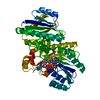
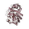
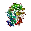
 Structure visualization
 Downloads & links
 Other downloads


 PDBj
PDBj
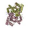
 Assembly
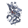

 Mass: 24.305 Da / Num. of mol.: 3 / Source method: obtained synthetically / Formula: Mg
 Mass: 550.499 Da / Num. of mol.: 2 / Source method: obtained synthetically / Formula: C21H22N6O10S
 Mass: 18.015 Da / Num. of mol.: 223 / Source method: isolated from a natural source / Formula: H2O
 Sample preparation
 Processing