 ムービー
ムービー コントローラー


+ データを開く

- 基本情報
基本情報
| 登録情報 | データベース: PDB / ID: 3u9x | ||||||
|---|---|---|---|---|---|---|---|
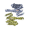
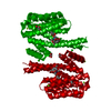
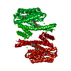
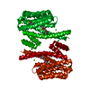
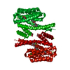
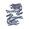
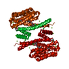
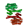
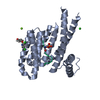
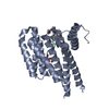
| タイトル | Covalent attachment of pyridoxal-phosphate derivatives to 14-3-3 proteins | ||||||
 要素 要素 | 14-3-3 protein sigma | ||||||
 キーワード キーワード |  SIGNALING PROTEIN/INHIBITOR / 14-3-3 (14-3-3タンパク質) / ADAPTER PROTEIN / PROTEIN-PROTEIN INTERACTION (タンパク質間相互作用) / N6-pyridoxal phosphate-L-lysine (PSI-MOD:128) / SIGNALING PROTEIN-INHIBITOR complex SIGNALING PROTEIN/INHIBITOR / 14-3-3 (14-3-3タンパク質) / ADAPTER PROTEIN / PROTEIN-PROTEIN INTERACTION (タンパク質間相互作用) / N6-pyridoxal phosphate-L-lysine (PSI-MOD:128) / SIGNALING PROTEIN-INHIBITOR complex | ||||||
| 機能・相同性 |  機能・相同性情報 機能・相同性情報regulation of epidermal cell division / protein kinase C inhibitor activity / positive regulation of epidermal cell differentiation / keratinocyte development / ケラチン / Regulation of localization of FOXO transcription factors / keratinocyte proliferation / phosphoserine residue binding / Activation of BAD and translocation to mitochondria / negative regulation of keratinocyte proliferation ...regulation of epidermal cell division / protein kinase C inhibitor activity / positive regulation of epidermal cell differentiation / keratinocyte development / ケラチン / Regulation of localization of FOXO transcription factors / keratinocyte proliferation / phosphoserine residue binding / Activation of BAD and translocation to mitochondria / negative regulation of keratinocyte proliferation / establishment of skin barrier / SARS-CoV-2 targets host intracellular signalling and regulatory pathways / Chk1/Chk2(Cds1) mediated inactivation of Cyclin B:Cdk1 complex / negative regulation of stem cell proliferation / SARS-CoV-1 targets host intracellular signalling and regulatory pathways / RHO GTPases activate PKNs / protein kinase A signaling / protein export from nucleus / negative regulation of innate immune response / protein sequestering activity / TP53 Regulates Transcription of Genes Involved in G2 Cell Cycle Arrest / release of cytochrome c from mitochondria / positive regulation of protein export from nucleus / stem cell proliferation / Translocation of SLC2A4 (GLUT4) to the plasma membrane / TP53 Regulates Metabolic Genes / negative regulation of protein kinase activity / negative regulation of cysteine-type endopeptidase activity involved in apoptotic process / intrinsic apoptotic signaling pathway in response to DNA damage / positive regulation of cell growth / regulation of cell cycle / cadherin binding / protein kinase binding / negative regulation of transcription by RNA polymerase II / シグナル伝達 / extracellular space / extracellular exosome / identical protein binding / 細胞核 / 細胞質基質 / 細胞質類似検索 - 分子機能 | ||||||
| 生物種 |  Homo sapiens (ヒト) Homo sapiens (ヒト) | ||||||
| 手法 | X線回折 / 分子置換 / 解像度: 1.8 Å | ||||||
 データ登録者 データ登録者 | Thiel, P. / Roeglin, L. / Kohlbacher, O. / Ottmann, C. | ||||||
 引用 引用 | ジャーナル: Proc.Natl.Acad.Sci.USA / 年: 2012 タイトル: Covalent attachment of pyridoxal-phosphate derivatives to 14-3-3 proteins. 著者: Roglin, L. / Thiel, P. / Kohlbacher, O. / Ottmann, C. | ||||||
| 履歴 |
|
- 構造の表示
構造の表示
| 構造ビューア | 分子: MolmilJmol/JSmol |
|---|
- ダウンロードとリンク
-ダウンロード
| PDBx/mmCIF形式 | 3u9x.cif.gz | 131.3 KB | 表示 | PDBx/mmCIF形式 |
|---|---|---|---|---|
| PDB形式 | pdb3u9x.ent.gz | 107.7 KB | 表示 | PDB形式 |
| PDBx/mmJSON形式 | 3u9x.json.gz | ツリー表示 | PDBx/mmJSON形式 | |
| その他 |  その他のダウンロード その他のダウンロード |
-検証レポート
| アーカイブディレクトリ | https://data.pdbj.org/pub/pdb/validation_reports/u9/3u9xftp://data.pdbj.org/pub/pdb/validation_reports/u9/3u9x | HTTPS FTP |
|---|
-関連構造データ
| 関連構造データ | |
|---|---|
| 類似構造データ |


-リンク
 PDBj
PDBj


- 集合体
集合体
| 登録構造単位 | 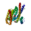
| ||||||||
|---|---|---|---|---|---|---|---|---|---|
| 1 | 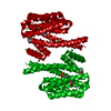
| ||||||||
| 単位格子 |
| ||||||||
| Components on special symmetry positions |
|
-要素
| #1: タンパク質 | 分子量: 26729.982 Da / 分子数: 1 / 断片: UNP residues 1-231 / 由来タイプ: 組換発現 / 由来: (組換発現) Homo sapiens (ヒト) / 遺伝子: HME1, NM_006142, SFN / プラスミド: PPROEX HTB / 発現宿主:  Escherichia coli (大腸菌) / 株 (発現宿主): ROSETTA (DE3) / 参照: UniProt: P31947 Escherichia coli (大腸菌) / 株 (発現宿主): ROSETTA (DE3) / 参照: UniProt: P31947 | ||||||
|---|---|---|---|---|---|---|---|
| #2: 化合物 |   分子量: 24.305 Da / 分子数: 2 / 由来タイプ: 合成 / 式: Mg 分子量: 24.305 Da / 分子数: 2 / 由来タイプ: 合成 / 式: Mg#3: 化合物 | 塩化物  分子量: 35.453 Da / 分子数: 3 / 由来タイプ: 合成 / 式: Cl 分子量: 35.453 Da / 分子数: 3 / 由来タイプ: 合成 / 式: Cl#4: 化合物 | ChemComp-GOL / | グリセリン  分子量: 92.094 Da / 分子数: 1 / 由来タイプ: 合成 / 式: C3H8O3 分子量: 92.094 Da / 分子数: 1 / 由来タイプ: 合成 / 式: C3H8O3#5: 水 | ChemComp-HOH / | 水 分子量: 18.015 Da / 分子数: 402 / 由来タイプ: 天然 / 式: H2O 分子量: 18.015 Da / 分子数: 402 / 由来タイプ: 天然 / 式: H2O |
-実験情報
-実験
| 実験 | 手法: X線回折 / 使用した結晶の数: 1 |
|---|
- 試料調製
試料調製
| 結晶 | マシュー密度: 2.7 Å3/Da / 溶媒含有率: 54.47 % |
|---|---|
| 結晶化 | 温度: 277 K / 手法: 蒸気拡散法, ハンギングドロップ法 / pH: 7.5 詳細: 0.095M HEPES Na, 26.6% PEG400, 0.1M Calcium chloride, 5% Glycerol, pH 7.5, VAPOR DIFFUSION, HANGING DROP, temperature 277K |
-データ収集
| 回折 | 平均測定温度: 100 K | ||||||||||||||||||||||||||||||||||||||||||||||||||||||||||||||||||||||||||||||||||||||||
|---|---|---|---|---|---|---|---|---|---|---|---|---|---|---|---|---|---|---|---|---|---|---|---|---|---|---|---|---|---|---|---|---|---|---|---|---|---|---|---|---|---|---|---|---|---|---|---|---|---|---|---|---|---|---|---|---|---|---|---|---|---|---|---|---|---|---|---|---|---|---|---|---|---|---|---|---|---|---|---|---|---|---|---|---|---|---|---|---|---|
| 放射光源 | 由来: 回転陽極 / タイプ: BRUKER AXS MICROSTAR / 波長: 1.5418 Å | ||||||||||||||||||||||||||||||||||||||||||||||||||||||||||||||||||||||||||||||||||||||||
| 検出器 | タイプ: MAR scanner 345 mm plate / 検出器: IMAGE PLATE / 日付: 2011年4月8日 / 詳細: mirrors | ||||||||||||||||||||||||||||||||||||||||||||||||||||||||||||||||||||||||||||||||||||||||
| 放射 | モノクロメーター: curved multilayer mirrors / プロトコル: SINGLE WAVELENGTH / 単色(M)・ラウエ(L): M / 散乱光タイプ: x-ray | ||||||||||||||||||||||||||||||||||||||||||||||||||||||||||||||||||||||||||||||||||||||||
| 放射波長 | 波長: 1.5418 Å / 相対比: 1 | ||||||||||||||||||||||||||||||||||||||||||||||||||||||||||||||||||||||||||||||||||||||||
| 反射 | 解像度: 1.8→45.54 Å / Num. all: 26966 / Num. obs: 26966 / % possible obs: 99 % / Observed criterion σ(F): -3 / Observed criterion σ(I): -3 / 冗長度: 5.8 % / Biso Wilson estimate: 22.755 Å2 / Rmerge(I) obs: 0.045 / Net I/σ(I): 35.76 | ||||||||||||||||||||||||||||||||||||||||||||||||||||||||||||||||||||||||||||||||||||||||
| 反射 シェル | Diffraction-ID: 1
|
-位相決定
| 位相決定 | 手法: 分子置換 | |||||||||
|---|---|---|---|---|---|---|---|---|---|---|
| Phasing MR | Rfactor: 28.82 / Model details: Phaser MODE: MR_AUTO
|
- 解析
解析
| ソフトウェア |
| ||||||||||||||||||||||||||||||||||||||||||||||||||||||||||||||||||||||||||||||||
|---|---|---|---|---|---|---|---|---|---|---|---|---|---|---|---|---|---|---|---|---|---|---|---|---|---|---|---|---|---|---|---|---|---|---|---|---|---|---|---|---|---|---|---|---|---|---|---|---|---|---|---|---|---|---|---|---|---|---|---|---|---|---|---|---|---|---|---|---|---|---|---|---|---|---|---|---|---|---|---|---|---|
| 精密化 | 構造決定の手法: 分子置換 / 解像度: 1.8→45.54 Å / Cor.coef. Fo:Fc: 0.969 / Cor.coef. Fo:Fc free: 0.945 / WRfactor Rfree: 0.1836 / WRfactor Rwork: 0.1272 / Occupancy max: 1 / Occupancy min: 0.21 / FOM work R set: 0.903 / SU B: 3.97 / SU ML: 0.057 / SU R Cruickshank DPI: 0.2372 / SU Rfree: 0.1087 / 交差検証法: THROUGHOUT / σ(F): 0 / ESU R Free: 0.109 / 立体化学のターゲット値: MAXIMUM LIKELIHOOD / 詳細: U VALUES : REFINED INDIVIDUALLY
| ||||||||||||||||||||||||||||||||||||||||||||||||||||||||||||||||||||||||||||||||
| 溶媒の処理 | イオンプローブ半径: 0.8 Å / 減衰半径: 0.8 Å / VDWプローブ半径: 1.4 Å / 溶媒モデル: MASK | ||||||||||||||||||||||||||||||||||||||||||||||||||||||||||||||||||||||||||||||||
| 原子変位パラメータ | Biso max: 60.16 Å2 / Biso mean: 17.6291 Å2 / Biso min: 3.74 Å2
| ||||||||||||||||||||||||||||||||||||||||||||||||||||||||||||||||||||||||||||||||
| 精密化ステップ | サイクル: LAST / 解像度: 1.8→45.54 Å
| ||||||||||||||||||||||||||||||||||||||||||||||||||||||||||||||||||||||||||||||||
| 拘束条件 |
| ||||||||||||||||||||||||||||||||||||||||||||||||||||||||||||||||||||||||||||||||
| LS精密化 シェル | 解像度: 1.8→1.847 Å / Total num. of bins used: 20
|
