 Movie
Movie Controller


+ Open data

- Basic information
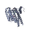
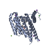
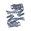
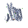
| Entry | Database: PDB / ID: 5n5t | ||||||
|---|---|---|---|---|---|---|---|
| Title | 14-3-3 sigma in complex with TAZ pS89 peptide and fragment NV2 | ||||||
 Components Components |
| ||||||
 Keywords Keywords |  PROTEIN BINDING / 14-3-3 / TAZ / fragment / FBDD PROTEIN BINDING / 14-3-3 / TAZ / fragment / FBDD | ||||||
| Function / homology |  Function and homology information Function and homology informationkidney morphogenesis / regulation of metanephric nephron tubule epithelial cell differentiation / Physiological factors / RUNX3 regulates YAP1-mediated transcription / mesenchymal cell differentiation / YAP1- and WWTR1 (TAZ)-stimulated gene expression / heart process / stem cell division / hippo signaling / tissue homeostasis ...kidney morphogenesis / regulation of metanephric nephron tubule epithelial cell differentiation / Physiological factors / RUNX3 regulates YAP1-mediated transcription / mesenchymal cell differentiation / YAP1- and WWTR1 (TAZ)-stimulated gene expression / heart process / stem cell division / hippo signaling / tissue homeostasis / EGR2 and SOX10-mediated initiation of Schwann cell myelination / glomerulus development / SMAD protein signal transduction / Signaling by Hippo / SCF-dependent proteasomal ubiquitin-dependent protein catabolic process / negative regulation of fat cell differentiation / regulation of epidermal cell division / protein kinase C inhibitor activity / positive regulation of epidermal cell differentiation / keratinocyte development / keratinization / RUNX2 regulates osteoblast differentiation / Regulation of localization of FOXO transcription factors / keratinocyte proliferation / cilium assembly / phosphoserine residue binding / Activation of BAD and translocation to mitochondria / negative regulation of keratinocyte proliferation / positive regulation of epithelial to mesenchymal transition / positive regulation of osteoblast differentiation / establishment of skin barrier / SARS-CoV-2 targets host intracellular signalling and regulatory pathways / Chk1/Chk2(Cds1) mediated inactivation of Cyclin B:Cdk1 complex / protein kinase A signaling / negative regulation of stem cell proliferation / SARS-CoV-1 targets host intracellular signalling and regulatory pathways / RHO GTPases activate PKNs / protein export from nucleus / negative regulation of innate immune response / protein sequestering activity / TP53 Regulates Transcription of Genes Involved in G2 Cell Cycle Arrest / release of cytochrome c from mitochondria / positive regulation of protein export from nucleus / negative regulation of protein phosphorylation / stem cell proliferation / Translocation of SLC2A4 (GLUT4) to the plasma membrane / TP53 Regulates Metabolic Genes / Downregulation of SMAD2/3:SMAD4 transcriptional activity / SMAD2/SMAD3:SMAD4 heterotrimer regulates transcription / negative regulation of protein kinase activity / multicellular organism growth / negative regulation of cysteine-type endopeptidase activity involved in apoptotic process / negative regulation of canonical Wnt signaling pathway / osteoblast differentiation / positive regulation of protein localization to nucleus / transcription corepressor activity / intrinsic apoptotic signaling pathway in response to DNA damage / positive regulation of cell growth / transcription regulator complex / transcription coactivator activity / protein ubiquitination / nuclear body / regulation of cell cycle / cadherin binding / positive regulation of cell population proliferation / regulation of DNA-templated transcription / protein kinase binding / negative regulation of transcription by RNA polymerase II / signal transduction / protein homodimerization activity / positive regulation of transcription by RNA polymerase II / extracellular space / extracellular exosome / nucleoplasm / identical protein binding / nucleus / plasma membrane / cytosol / cytoplasmSimilarity search - Function | ||||||
| Biological species |  Homo sapiens (human) Homo sapiens (human) | ||||||
| Method | X-RAY DIFFRACTION / MOLECULAR REPLACEMENT / Resolution: 1.8 Å | ||||||
 Authors Authors | Sijbesma, E. / Leysen, S. / Ottmann, C. | ||||||
 Citation Citation | Journal: Biochemistry / Year: 2017 Title: Identification of Two Secondary Ligand Binding Sites in 14-3-3 Proteins Using Fragment Screening. Authors: Sijbesma, E. / Skora, L. / Leysen, S. / Brunsveld, L. / Koch, U. / Nussbaumer, P. / Jahnke, W. / Ottmann, C. | ||||||
| History |
|
- Structure visualization
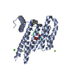
| Structure viewer | Molecule: MolmilJmol/JSmol |
|---|
- Downloads & links
-Download
| PDBx/mmCIF format | 5n5t.cif.gz | 114.1 KB | Display | PDBx/mmCIF format |
|---|---|---|---|---|
| PDB format | pdb5n5t.ent.gz | 88.5 KB | Display | PDB format |
| PDBx/mmJSON format | 5n5t.json.gz | Tree view | PDBx/mmJSON format | |
| Others |  Other downloads Other downloads |
-Validation report
| Arichive directory | https://data.pdbj.org/pub/pdb/validation_reports/n5/5n5tftp://data.pdbj.org/pub/pdb/validation_reports/n5/5n5t | HTTPS FTP |
|---|
-Related structure data
| Related structure data | 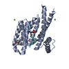 5n5rC 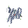 5n5wC 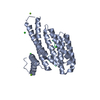 5n75C 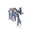 3mhrS S: Starting model for refinement C: citing same article ( |
|---|---|
| Similar structure data |


-Links
 PDBj
PDBj


- Assembly
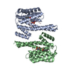
| Deposited unit | 
| ||||||||
|---|---|---|---|---|---|---|---|---|---|
| 1 | 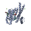
| ||||||||
| Unit cell |
|
-Components
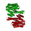
-Protein / Protein/peptide , 2 types, 2 molecules AP
| #1: Protein | Mass: 26542.914 Da / Num. of mol.: 1 Source method: isolated from a genetically manipulated source Source: (gene. exp.) Homo sapiens (human) / Gene: SFN, HME1 / Production host:  Escherichia coli (E. coli) / References: UniProt: P31947 Escherichia coli (E. coli) / References: UniProt: P31947 |
|---|---|
| #2: Protein/peptide | Mass: 865.803 Da / Num. of mol.: 1 / Source method: obtained synthetically / Source: (synth.) Homo sapiens (human) / References: UniProt: Q9GZV5*PLUS |
-Non-polymers , 4 types, 252 molecules 


| #3: Chemical |  Mass: 24.305 Da / Num. of mol.: 2 / Source method: obtained synthetically / Formula: Mg Mass: 24.305 Da / Num. of mol.: 2 / Source method: obtained synthetically / Formula: Mg#4: Chemical | ChemComp-CL / | Chloride Mass: 35.453 Da / Num. of mol.: 1 / Source method: obtained synthetically / Formula: Cl Mass: 35.453 Da / Num. of mol.: 1 / Source method: obtained synthetically / Formula: Cl#5: Chemical | ChemComp-8OB / |  Mass: 256.085 Da / Num. of mol.: 1 / Source method: obtained synthetically / Formula: C11H7Cl2NO2 Mass: 256.085 Da / Num. of mol.: 1 / Source method: obtained synthetically / Formula: C11H7Cl2NO2#6: Water | ChemComp-HOH / | WaterMass: 18.015 Da / Num. of mol.: 248 / Source method: isolated from a natural source / Formula: H2O |
|---|
-Experimental details
-Experiment
| Experiment | Method: X-RAY DIFFRACTION / Number of used crystals: 1 |
|---|
- Sample preparation
| Crystal | Density Matthews: 2.61 Å3/Da / Density % sol: 52.92 % |
|---|---|
| Crystal grow | Temperature: 277 K / Method: vapor diffusion, sitting drop / pH: 7.1 Details: 0.095 M Na-HEPES pH 7.1, 28% PEG400, 0.19 M Calcium chloride, 5% Glycerol |
-Data collection
| Diffraction | Mean temperature: 100 K | |||||||||||||||||||||
|---|---|---|---|---|---|---|---|---|---|---|---|---|---|---|---|---|---|---|---|---|---|---|
| Diffraction source | Source: ROTATING ANODE / Type: RIGAKU MICROMAX-003 / Wavelength: 1.54 Å | |||||||||||||||||||||
| Detector | Type: DECTRIS PILATUS 200K / Detector: PIXEL / Date: Jun 11, 2015 | |||||||||||||||||||||
| Radiation | Protocol: SINGLE WAVELENGTH / Monochromatic (M) / Laue (L): M / Scattering type: x-ray | |||||||||||||||||||||
| Radiation wavelength | Wavelength: 1.54 Å / Relative weight: 1 | |||||||||||||||||||||
| Reflection | Resolution: 1.8→65.96 Å / Num. obs: 26972 / % possible obs: 99.8 % / Redundancy: 6.2 % / Biso Wilson estimate: 12.35 Å2 / CC1/2: 1 / Rmerge(I) obs: 0.034 / Rpim(I) all: 0.015 / Rrim(I) all: 0.037 / Net I/σ(I): 42.1 / Num. measured all: 167853 / Scaling rejects: 0 | |||||||||||||||||||||
| Reflection shell | Diffraction-ID: 1
|
- Processing
| Software |
| ||||||||||||||||||||||||||||||||||||||||||||||||||||||||||||||||||||||
|---|---|---|---|---|---|---|---|---|---|---|---|---|---|---|---|---|---|---|---|---|---|---|---|---|---|---|---|---|---|---|---|---|---|---|---|---|---|---|---|---|---|---|---|---|---|---|---|---|---|---|---|---|---|---|---|---|---|---|---|---|---|---|---|---|---|---|---|---|---|---|---|
| Refinement | Method to determine structure: MOLECULAR REPLACEMENT Starting model: 3MHR Resolution: 1.8→34.154 Å / SU ML: 0.12 / Cross valid method: FREE R-VALUE / σ(F): 1.34 / Phase error: 16.27
| ||||||||||||||||||||||||||||||||||||||||||||||||||||||||||||||||||||||
| Solvent computation | Shrinkage radii: 0.9 Å / VDW probe radii: 1.11 Å | ||||||||||||||||||||||||||||||||||||||||||||||||||||||||||||||||||||||
| Displacement parameters | Biso max: 75.44 Å2 / Biso mean: 15.6431 Å2 / Biso min: 4.71 Å2 | ||||||||||||||||||||||||||||||||||||||||||||||||||||||||||||||||||||||
| Refinement step | Cycle: final / Resolution: 1.8→34.154 Å
| ||||||||||||||||||||||||||||||||||||||||||||||||||||||||||||||||||||||
| Refine LS restraints |
| ||||||||||||||||||||||||||||||||||||||||||||||||||||||||||||||||||||||
| LS refinement shell | Refine-ID: X-RAY DIFFRACTION / Rfactor Rfree error: 0 / Total num. of bins used: 9
|
