 Movie
Movie Controller

[English] 日本語
 Yorodumi
- PDB-3u8g: Crystal structure of the complex of Dihydrodipicolinate synthase ... -
+ Open data

- Basic information
| Entry | Database: PDB / ID: 3u8g | ||||||
|---|---|---|---|---|---|---|---|
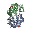
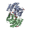
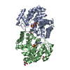
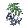
| Title | Crystal structure of the complex of Dihydrodipicolinate synthase from Acinetobacter baumannii with Oxalic acid at 1.80 A resolution | ||||||
 Components Components | Dihydrodipicolinate synthase | ||||||
 Keywords Keywords | LYASE / DHDPS / Lysine biosynthesis / Oxalic acid / TIM Barrel | ||||||
| Function / homology |  Function and homology information4-hydroxy-2-oxoglutarate aldolase activity / 4-hydroxy-tetrahydrodipicolinate synthase / 4-hydroxy-tetrahydrodipicolinate synthase activity / glyoxylate catabolic process / diaminopimelate biosynthetic process / lysine biosynthetic process via diaminopimelate / metal ion binding / cytosol Function and homology information4-hydroxy-2-oxoglutarate aldolase activity / 4-hydroxy-tetrahydrodipicolinate synthase / 4-hydroxy-tetrahydrodipicolinate synthase activity / glyoxylate catabolic process / diaminopimelate biosynthetic process / lysine biosynthetic process via diaminopimelate / metal ion binding / cytosolSimilarity search - Function | ||||||
| Biological species |  Acinetobacter baumannii (bacteria) Acinetobacter baumannii (bacteria) | ||||||
| Method | X-RAY DIFFRACTION / SYNCHROTRON / MOLECULAR REPLACEMENT / Resolution: 1.8 Å | ||||||
 Authors Authors | Kumar, M. / Kaushik, S. / Bhushan, A. / Sinha, M. / Kaur, P. / Sharma, S. / Singh, T.P. | ||||||
 Citation Citation | Journal: To be Published Title: Crystal structure of the complex of Dihydrodipicolinate synthase from Acinetobacter baumannii with Oxalic acid at 1.80 A resolution Authors: Kumar, M. / Kaushik, S. / Bhushan, A. / Sinha, M. / Kaur, P. / Sharma, S. / Singh, T.P. | ||||||
| History |
|
- Structure visualization
| Structure viewer | Molecule: MolmilJmol/JSmol |
|---|
- Downloads & links
-Download
| PDBx/mmCIF format | 3u8g.cif.gz | 132.1 KB | Display | PDBx/mmCIF format |
|---|---|---|---|---|
| PDB format | pdb3u8g.ent.gz | 102 KB | Display | PDB format |
| PDBx/mmJSON format | 3u8g.json.gz | Tree view | PDBx/mmJSON format | |
| Others |  Other downloads Other downloads |
-Validation report
| Arichive directory | https://data.pdbj.org/pub/pdb/validation_reports/u8/3u8gftp://data.pdbj.org/pub/pdb/validation_reports/u8/3u8g | HTTPS FTP |
|---|
-Related structure data
| Related structure data |  3rk8S S: Starting model for refinement |
|---|---|
| Similar structure data |


-Links
 PDBj
PDBj- Assembly
| Deposited unit | 
| ||||||||
|---|---|---|---|---|---|---|---|---|---|
| 1 |
| ||||||||
| Unit cell |
|
-Components
| #1: Protein | / DHDPS Mass: 31279.842 Da / Num. of mol.: 2 / Fragment: UNP residues 7-297 Source method: isolated from a genetically manipulated source Source: (gene. exp.) Acinetobacter baumannii (bacteria) / Strain: ATCC 19606 / Gene: dhdps, HMPREF0010_03414 / Plasmid: pRESTa / Production host: Escherichia coli (E. coli) / Strain (production host): Bl21(de3) / References: UniProt: D0CFC3, dihydrodipicolinate synthase#2: Chemical | Oxalate  Mass: 88.019 Da / Num. of mol.: 2 / Source method: obtained synthetically / Formula: C2O4 Mass: 88.019 Da / Num. of mol.: 2 / Source method: obtained synthetically / Formula: C2O4#3: Water | ChemComp-HOH / | Water Mass: 18.015 Da / Num. of mol.: 621 / Source method: isolated from a natural source / Formula: H2O Mass: 18.015 Da / Num. of mol.: 621 / Source method: isolated from a natural source / Formula: H2O |
|---|
-Experimental details
-Experiment
| Experiment | Method: X-RAY DIFFRACTION / Number of used crystals: 1 |
|---|
- Sample preparation
| Crystal | Density Matthews: 2.37 Å3/Da / Density % sol: 48.01 % |
|---|---|
| Crystal grow | Temperature: 298 K / Method: vapor diffusion, hanging drop / pH: 6.2 Details: 0.1M MgCl2, PEG 3350, pH 6.2, VAPOR DIFFUSION, HANGING DROP, temperature 298K |
-Data collection
| Diffraction | Mean temperature: 77 K |
|---|---|
| Diffraction source | Source: SYNCHROTRON / Site: ESRF  / Beamline: BM14 / Wavelength: 0.97 Å / Beamline: BM14 / Wavelength: 0.97 Å |
| Detector | Type: MARRESEARCH / Detector: CCD / Date: Sep 22, 2011 / Details: MIRROR |
| Radiation | Monochromator: Graphite / Protocol: SINGLE WAVELENGTH / Monochromatic (M) / Laue (L): M / Scattering type: x-ray |
| Radiation wavelength | Wavelength: 0.97 Å / Relative weight: 1 |
| Reflection | Resolution: 1.8→61.2 Å / Num. all: 51034 / Num. obs: 51034 / % possible obs: 99 % / Observed criterion σ(F): 0 / Observed criterion σ(I): 0 / Rsym value: 0.098 / Net I/σ(I): 19 |
| Reflection shell | Resolution: 1.8→1.83 Å / Mean I/σ(I) obs: 2.9 / Rsym value: 0.448 / % possible all: 100 |
- Processing
| Software |
| |||||||||||||||||||||||||||||||||||||||||||||||||||||||||||||||||
|---|---|---|---|---|---|---|---|---|---|---|---|---|---|---|---|---|---|---|---|---|---|---|---|---|---|---|---|---|---|---|---|---|---|---|---|---|---|---|---|---|---|---|---|---|---|---|---|---|---|---|---|---|---|---|---|---|---|---|---|---|---|---|---|---|---|---|
| Refinement | Method to determine structure: MOLECULAR REPLACEMENT Starting model: 3RK8 Resolution: 1.8→47.09 Å / Cor.coef. Fo:Fc: 0.954 / Cor.coef. Fo:Fc free: 0.936 / SU B: 2.349 / SU ML: 0.074 / Cross valid method: THROUGHOUT / σ(F): 0 / σ(I): 0 / ESU R Free: 0.122 / Stereochemistry target values: MAXIMUM LIKELIHOOD / Details: HYDROGENS HAVE BEEN ADDED IN THE RIDING POSITIONS
| |||||||||||||||||||||||||||||||||||||||||||||||||||||||||||||||||
| Solvent computation | Ion probe radii: 0.8 Å / Shrinkage radii: 0.8 Å / VDW probe radii: 1.4 Å / Solvent model: MASK | |||||||||||||||||||||||||||||||||||||||||||||||||||||||||||||||||
| Displacement parameters | Biso mean: 16.857 Å2
| |||||||||||||||||||||||||||||||||||||||||||||||||||||||||||||||||
| Refinement step | Cycle: LAST / Resolution: 1.8→47.09 Å
| |||||||||||||||||||||||||||||||||||||||||||||||||||||||||||||||||
| Refine LS restraints |
| |||||||||||||||||||||||||||||||||||||||||||||||||||||||||||||||||
| LS refinement shell | Resolution: 1.798→1.845 Å / Total num. of bins used: 20
|
