 Movie
Movie Controller

[English] 日本語
 Yorodumi
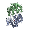
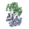
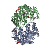
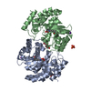
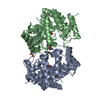
- PDB-3pue: Crystal structure of the complex of Dhydrodipicolinate synthase f... -
+ Open data

- Basic information
| Entry | Database: PDB / ID: 3pue | ||||||
|---|---|---|---|---|---|---|---|
| Title | Crystal structure of the complex of Dhydrodipicolinate synthase from Acinetobacter baumannii with lysine at 2.6A resolution | ||||||
 Components Components | Dihydrodipicolinate synthase | ||||||
 Keywords Keywords | LYASE / TIM barrel / beta-alpha-barrel / Diaminopimelate biosynthesis / Lysine biosynthesis | ||||||
| Function / homology |  Function and homology information4-hydroxy-2-oxoglutarate aldolase activity / 4-hydroxy-tetrahydrodipicolinate synthase / 4-hydroxy-tetrahydrodipicolinate synthase activity / glyoxylate catabolic process / diaminopimelate biosynthetic process / lysine biosynthetic process via diaminopimelate / metal ion binding / cytosol Function and homology information4-hydroxy-2-oxoglutarate aldolase activity / 4-hydroxy-tetrahydrodipicolinate synthase / 4-hydroxy-tetrahydrodipicolinate synthase activity / glyoxylate catabolic process / diaminopimelate biosynthetic process / lysine biosynthetic process via diaminopimelate / metal ion binding / cytosolSimilarity search - Function | ||||||
| Biological species |  Acinetobacter baumannii (bacteria) Acinetobacter baumannii (bacteria) | ||||||
| Method | X-RAY DIFFRACTION / SYNCHROTRON / MOLECULAR REPLACEMENT / Resolution: 2.6 Å | ||||||
 Authors Authors | Jithesh, O. / Yamini, S. / Kaur, N. / Gautam, A. / Tewari, R. / Kushwaha, G.S. / Kaur, P. / Srinivasan, A. / Sharma, S. / Singh, T.P. | ||||||
 Citation Citation | Journal: To be Published Title: Crystal structure of the complex of Dhydrodipicolinate synthase from Acinetobacter baumannii with lysine at 2.6A resolution Authors: Jithesh, O. / Yamini, S. / Kaur, N. / Gautam, A. / Tewari, R. / Kushwaha, G.S. / Kaur, P. / Srinivasan, A. / Sharma, S. / Singh, T.P. | ||||||
| History |
|
- Structure visualization
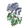
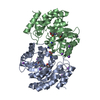
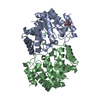
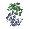
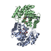
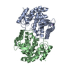
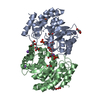
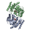
| Structure viewer | Molecule: MolmilJmol/JSmol |
|---|
- Downloads & links
-Download
| PDBx/mmCIF format | 3pue.cif.gz | 122.6 KB | Display | PDBx/mmCIF format |
|---|---|---|---|---|
| PDB format | pdb3pue.ent.gz | 95.9 KB | Display | PDB format |
| PDBx/mmJSON format | 3pue.json.gz | Tree view | PDBx/mmJSON format | |
| Others |  Other downloads Other downloads |
-Validation report
| Arichive directory | https://data.pdbj.org/pub/pdb/validation_reports/pu/3pueftp://data.pdbj.org/pub/pdb/validation_reports/pu/3pue | HTTPS FTP |
|---|
-Related structure data
| Related structure data |  3noeS S: Starting model for refinement |
|---|---|
| Similar structure data |


-Links
 PDBj
PDBj- Assembly
| Deposited unit | 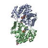
| ||||||||
|---|---|---|---|---|---|---|---|---|---|
| 1 |
| ||||||||
| Unit cell |
|
-Components
| #1: Protein | Mass: 31279.842 Da / Num. of mol.: 2 / Fragment: DHDPS, residues in UNP 7-297 Source method: isolated from a genetically manipulated source Source: (gene. exp.) Acinetobacter baumannii (bacteria) / Strain: ATCC 19606 / Gene: DHDPS, HMPREF0010_03414 / Plasmid: pRSETa / Production host: Escherichia coli (E. coli) / Strain (production host): BL21(DE3) / References: UniProt: D0CFC3, dihydrodipicolinate synthase#2: Chemical | Sulfate  Mass: 96.063 Da / Num. of mol.: 3 / Source method: obtained synthetically / Formula: SO4 Mass: 96.063 Da / Num. of mol.: 3 / Source method: obtained synthetically / Formula: SO4#3: Chemical | ChemComp-LYS / | Lysine  Type: L-peptide linking / Mass: 147.195 Da / Num. of mol.: 1 / Source method: obtained synthetically / Formula: C6H15N2O2 Type: L-peptide linking / Mass: 147.195 Da / Num. of mol.: 1 / Source method: obtained synthetically / Formula: C6H15N2O2#4: Chemical | Glycerol  Mass: 92.094 Da / Num. of mol.: 3 / Source method: obtained synthetically / Formula: C3H8O3 Mass: 92.094 Da / Num. of mol.: 3 / Source method: obtained synthetically / Formula: C3H8O3#5: Water | ChemComp-HOH / | Water Mass: 18.015 Da / Num. of mol.: 164 / Source method: isolated from a natural source / Formula: H2O Mass: 18.015 Da / Num. of mol.: 164 / Source method: isolated from a natural source / Formula: H2O |
|---|
-Experimental details
-Experiment
| Experiment | Method: X-RAY DIFFRACTION / Number of used crystals: 1 |
|---|
- Sample preparation
| Crystal | Density Matthews: 2.72 Å3/Da / Density % sol: 54.78 % |
|---|---|
| Crystal grow | Temperature: 298 K / Method: vapor diffusion / pH: 5.6 Details: 1.8M ammonium sulphate, 0.1M bis-tris, pH 5.6, VAPOR DIFFUSION, temperature 298K |
-Data collection
| Diffraction | Mean temperature: 77 K |
|---|---|
| Diffraction source | Source: SYNCHROTRON / Site: ESRF  / Beamline: BM14 / Wavelength: 0.97 Å / Beamline: BM14 / Wavelength: 0.97 Å |
| Detector | Type: MARRESEARCH / Detector: CCD / Date: Oct 21, 2010 / Details: MIRROR |
| Radiation | Monochromator: GRAPHITE / Protocol: SINGLE WAVELENGTH / Monochromatic (M) / Laue (L): M / Scattering type: x-ray |
| Radiation wavelength | Wavelength: 0.97 Å / Relative weight: 1 |
| Reflection | Resolution: 2.6→84.5 Å / Num. all: 20253 / Num. obs: 20253 / % possible obs: 99.7 % / Observed criterion σ(F): 0 / Observed criterion σ(I): 0 / Biso Wilson estimate: 49.4 Å2 / Rsym value: 0.082 / Net I/σ(I): 15.1 |
| Reflection shell | Resolution: 2.6→2.64 Å / Mean I/σ(I) obs: 2.3 / Rsym value: 0.378 / % possible all: 97 |
- Processing
| Software |
| ||||||||||||||||||||||||||||||||||||||||||||||||||||||||||||||||||||||||||||||||
|---|---|---|---|---|---|---|---|---|---|---|---|---|---|---|---|---|---|---|---|---|---|---|---|---|---|---|---|---|---|---|---|---|---|---|---|---|---|---|---|---|---|---|---|---|---|---|---|---|---|---|---|---|---|---|---|---|---|---|---|---|---|---|---|---|---|---|---|---|---|---|---|---|---|---|---|---|---|---|---|---|---|
| Refinement | Method to determine structure: MOLECULAR REPLACEMENT Starting model: 3NOE Resolution: 2.6→48.8 Å / Rfactor Rfree error: 0.008 / Data cutoff high absF: 6550898.97 / Data cutoff low absF: 0 / Isotropic thermal model: RESTRAINED / Cross valid method: THROUGHOUT / σ(F): 0 / Stereochemistry target values: MAXIMUM LIKELIHOOD
| ||||||||||||||||||||||||||||||||||||||||||||||||||||||||||||||||||||||||||||||||
| Solvent computation | Solvent model: FLAT MODEL / Bsol: 38.733 Å2 / ksol: 0.392944 e/Å3 | ||||||||||||||||||||||||||||||||||||||||||||||||||||||||||||||||||||||||||||||||
| Displacement parameters | Biso mean: 34.8 Å2
| ||||||||||||||||||||||||||||||||||||||||||||||||||||||||||||||||||||||||||||||||
| Refine analyze |
| ||||||||||||||||||||||||||||||||||||||||||||||||||||||||||||||||||||||||||||||||
| Refinement step | Cycle: LAST / Resolution: 2.6→48.8 Å
| ||||||||||||||||||||||||||||||||||||||||||||||||||||||||||||||||||||||||||||||||
| Refine LS restraints |
| ||||||||||||||||||||||||||||||||||||||||||||||||||||||||||||||||||||||||||||||||
| LS refinement shell | Resolution: 2.6→2.76 Å / Rfactor Rfree error: 0.024 / Total num. of bins used: 6
| ||||||||||||||||||||||||||||||||||||||||||||||||||||||||||||||||||||||||||||||||
| Xplor file |
|
