 Movie
Movie Controller

[English] 日本語
 Yorodumi
- PDB-3daq: Crystal structure of dihydrodipicolinate synthase from methicilli... -
+ Open data

- Basic information
| Entry | Database: PDB / ID: 3daq | ||||||
|---|---|---|---|---|---|---|---|
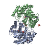
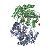
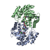
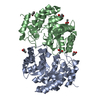
| Title | Crystal structure of dihydrodipicolinate synthase from methicillin-resistant Staphylococcus aureus | ||||||
 Components Components | Dihydrodipicolinate synthase | ||||||
 Keywords Keywords | LYASE / dihydrodipicolinate synthase / lysine biosynthesis / Amino-acid biosynthesis / Diaminopimelate biosynthesis / Schiff base | ||||||
| Function / homology |  Function and homology information4-hydroxy-tetrahydrodipicolinate synthase / 4-hydroxy-tetrahydrodipicolinate synthase activity / diaminopimelate biosynthetic process / lysine biosynthetic process via diaminopimelate / cytoplasm Function and homology information4-hydroxy-tetrahydrodipicolinate synthase / 4-hydroxy-tetrahydrodipicolinate synthase activity / diaminopimelate biosynthetic process / lysine biosynthetic process via diaminopimelate / cytoplasmSimilarity search - Function | ||||||
| Biological species |   Staphylococcus aureus (bacteria) Staphylococcus aureus (bacteria) | ||||||
| Method | X-RAY DIFFRACTION / SYNCHROTRON / MOLECULAR REPLACEMENT / Resolution: 1.45 Å | ||||||
 Authors Authors | Dobson, R.C.J. / Burgess, B.R. / Jameson, G.B. / Gerrard, J.A. / Parker, M.W. / Perugini, M.A. | ||||||
 Citation Citation | Journal: J.Biol.Chem. / Year: 2008 Title: Structure and evolution of a novel dimeric enzyme from a clinically-important bacterial pathogen. Authors: Burgess, B.R. / Dobson, R.C.J. / Bailey, M.F. / Atkinson, S.C. / Griffin, M.D.W. / Jameson, G.B. / Parker, M.W. / Gerrard, J.A. / Perugini, M.A. | ||||||
| History |
|
- Structure visualization
| Structure viewer | Molecule: MolmilJmol/JSmol |
|---|
- Downloads & links
-Download
| PDBx/mmCIF format | 3daq.cif.gz | 510 KB | Display | PDBx/mmCIF format |
|---|---|---|---|---|
| PDB format | pdb3daq.ent.gz | 418.7 KB | Display | PDB format |
| PDBx/mmJSON format | 3daq.json.gz | Tree view | PDBx/mmJSON format | |
| Others |  Other downloads Other downloads |
-Validation report
| Arichive directory | https://data.pdbj.org/pub/pdb/validation_reports/da/3daqftp://data.pdbj.org/pub/pdb/validation_reports/da/3daq | HTTPS FTP |
|---|
-Related structure data
| Related structure data |  1yxcS S: Starting model for refinement |
|---|---|
| Similar structure data |


-Links
 PDBj
PDBj- Assembly
| Deposited unit | 
| ||||||||||||||||||||||||||||||||||||||||||||||||||||||||||||||||||||||||||||||||||||||||||||||||||||||||||||||||||||||||||||||||||||||||||||||||||||||||||||||||||||||||||||||||||||||||||
|---|---|---|---|---|---|---|---|---|---|---|---|---|---|---|---|---|---|---|---|---|---|---|---|---|---|---|---|---|---|---|---|---|---|---|---|---|---|---|---|---|---|---|---|---|---|---|---|---|---|---|---|---|---|---|---|---|---|---|---|---|---|---|---|---|---|---|---|---|---|---|---|---|---|---|---|---|---|---|---|---|---|---|---|---|---|---|---|---|---|---|---|---|---|---|---|---|---|---|---|---|---|---|---|---|---|---|---|---|---|---|---|---|---|---|---|---|---|---|---|---|---|---|---|---|---|---|---|---|---|---|---|---|---|---|---|---|---|---|---|---|---|---|---|---|---|---|---|---|---|---|---|---|---|---|---|---|---|---|---|---|---|---|---|---|---|---|---|---|---|---|---|---|---|---|---|---|---|---|---|---|---|---|---|---|---|---|---|
| 1 | 
| ||||||||||||||||||||||||||||||||||||||||||||||||||||||||||||||||||||||||||||||||||||||||||||||||||||||||||||||||||||||||||||||||||||||||||||||||||||||||||||||||||||||||||||||||||||||||||
| 2 | 
| ||||||||||||||||||||||||||||||||||||||||||||||||||||||||||||||||||||||||||||||||||||||||||||||||||||||||||||||||||||||||||||||||||||||||||||||||||||||||||||||||||||||||||||||||||||||||||
| Unit cell |
| ||||||||||||||||||||||||||||||||||||||||||||||||||||||||||||||||||||||||||||||||||||||||||||||||||||||||||||||||||||||||||||||||||||||||||||||||||||||||||||||||||||||||||||||||||||||||||
| Noncrystallographic symmetry (NCS) | NCS domain:
NCS domain segments: Component-ID: 1
NCS ensembles :
|
-Components
| #1: Protein | / DHDPS Mass: 32135.295 Da / Num. of mol.: 4 / Fragment: residues 2-293 Source method: isolated from a genetically manipulated source Source: (gene. exp.) Staphylococcus aureus (bacteria) / Gene: dapA, SAR1407 / Plasmid: pET11a / Production host: Escherichia coli (E. coli) / Strain (production host): BL21 (DE3) / References: UniProt: Q6GH13, dihydrodipicolinate synthase#2: Chemical | ChemComp-CL / Chloride  Mass: 35.453 Da / Num. of mol.: 4 / Source method: obtained synthetically / Formula: Cl Mass: 35.453 Da / Num. of mol.: 4 / Source method: obtained synthetically / Formula: Cl#3: Chemical | ChemComp-GOL / Glycerol  Mass: 92.094 Da / Num. of mol.: 9 / Source method: obtained synthetically / Formula: C3H8O3 Mass: 92.094 Da / Num. of mol.: 9 / Source method: obtained synthetically / Formula: C3H8O3#4: Water | ChemComp-HOH / | Water Mass: 18.015 Da / Num. of mol.: 1795 / Source method: isolated from a natural source / Formula: H2O Mass: 18.015 Da / Num. of mol.: 1795 / Source method: isolated from a natural source / Formula: H2O |
|---|
-Experimental details
-Experiment
| Experiment | Method: X-RAY DIFFRACTION / Number of used crystals: 1 |
|---|
- Sample preparation
| Crystal | Density Matthews: 2.36 Å3/Da / Density % sol: 47.96 % |
|---|---|
| Crystal grow | Temperature: 281 K / Method: vapor diffusion, hanging drop / pH: 4.9 Details: 16.7% PEG6000, NaF 135mM, LiCl 296mM, NaAC 100mM, pH 4.9, VAPOR DIFFUSION, HANGING DROP, temperature 281K |
-Data collection
| Diffraction | Mean temperature: 100 K |
|---|---|
| Diffraction source | Source: SYNCHROTRON / Site: Australian Synchrotron  / Beamline: MX1 / Wavelength: 0.9536 Å / Beamline: MX1 / Wavelength: 0.9536 Å |
| Detector | Type: ADSC QUANTUM 210 / Detector: CCD / Date: Apr 10, 2008 |
| Radiation | Protocol: SINGLE WAVELENGTH / Monochromatic (M) / Laue (L): M / Scattering type: x-ray |
| Radiation wavelength | Wavelength: 0.9536 Å / Relative weight: 1 |
| Reflection | Resolution: 1.45→30.25 Å / Num. obs: 190869 / % possible obs: 91.4 % / Observed criterion σ(F): 2 / Observed criterion σ(I): 2 / Redundancy: 3.9 % / Biso Wilson estimate: 9.84 Å2 / Rmerge(I) obs: 0.057 / Rsym value: 0.057 / Net I/σ(I): 18.8 |
| Reflection shell | Resolution: 1.45→1.53 Å / Redundancy: 3.9 % / Rmerge(I) obs: 0.147 / Mean I/σ(I) obs: 8.4 / Num. unique all: 21900 / Rsym value: 0.147 / % possible all: 71.9 |
- Processing
| Software |
| |||||||||||||||||||||||||||||||||||||||||||||||||||||||||||||||||||||||||||||||||||||||||||||||||||||||||||||||||||||||||||||||||||||||||||||||||||||||||||||||||||||||||||||||||||||||||||||||||||||||
|---|---|---|---|---|---|---|---|---|---|---|---|---|---|---|---|---|---|---|---|---|---|---|---|---|---|---|---|---|---|---|---|---|---|---|---|---|---|---|---|---|---|---|---|---|---|---|---|---|---|---|---|---|---|---|---|---|---|---|---|---|---|---|---|---|---|---|---|---|---|---|---|---|---|---|---|---|---|---|---|---|---|---|---|---|---|---|---|---|---|---|---|---|---|---|---|---|---|---|---|---|---|---|---|---|---|---|---|---|---|---|---|---|---|---|---|---|---|---|---|---|---|---|---|---|---|---|---|---|---|---|---|---|---|---|---|---|---|---|---|---|---|---|---|---|---|---|---|---|---|---|---|---|---|---|---|---|---|---|---|---|---|---|---|---|---|---|---|---|---|---|---|---|---|---|---|---|---|---|---|---|---|---|---|---|---|---|---|---|---|---|---|---|---|---|---|---|---|---|---|---|
| Refinement | Method to determine structure: MOLECULAR REPLACEMENT Starting model: 1yxc Resolution: 1.45→30.25 Å / Cor.coef. Fo:Fc: 0.97 / Cor.coef. Fo:Fc free: 0.958 / SU B: 1.84 / SU ML: 0.033 / Cross valid method: THROUGHOUT / ESU R: 0.071 / ESU R Free: 0.058 / Stereochemistry target values: MAXIMUM LIKELIHOOD / Details: HYDROGENS HAVE BEEN ADDED IN THE RIDING POSITIONS
| |||||||||||||||||||||||||||||||||||||||||||||||||||||||||||||||||||||||||||||||||||||||||||||||||||||||||||||||||||||||||||||||||||||||||||||||||||||||||||||||||||||||||||||||||||||||||||||||||||||||
| Solvent computation | Ion probe radii: 0.8 Å / Shrinkage radii: 0.8 Å / VDW probe radii: 1.2 Å / Solvent model: BABINET MODEL WITH MASK | |||||||||||||||||||||||||||||||||||||||||||||||||||||||||||||||||||||||||||||||||||||||||||||||||||||||||||||||||||||||||||||||||||||||||||||||||||||||||||||||||||||||||||||||||||||||||||||||||||||||
| Displacement parameters | Biso mean: 8.073 Å2
| |||||||||||||||||||||||||||||||||||||||||||||||||||||||||||||||||||||||||||||||||||||||||||||||||||||||||||||||||||||||||||||||||||||||||||||||||||||||||||||||||||||||||||||||||||||||||||||||||||||||
| Refinement step | Cycle: LAST / Resolution: 1.45→30.25 Å
| |||||||||||||||||||||||||||||||||||||||||||||||||||||||||||||||||||||||||||||||||||||||||||||||||||||||||||||||||||||||||||||||||||||||||||||||||||||||||||||||||||||||||||||||||||||||||||||||||||||||
| Refine LS restraints |
| |||||||||||||||||||||||||||||||||||||||||||||||||||||||||||||||||||||||||||||||||||||||||||||||||||||||||||||||||||||||||||||||||||||||||||||||||||||||||||||||||||||||||||||||||||||||||||||||||||||||
| Refine LS restraints NCS | Refine-ID: X-RAY DIFFRACTION
|