 Movie
Movie Controller


+ Open data

- Basic information
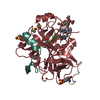
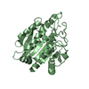
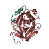
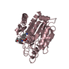
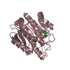
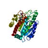
| Entry | Database: PDB / ID: 3u3k | ||||||
|---|---|---|---|---|---|---|---|
| Title | Crystal structure of hSULT1A1 bound to PAP and 2-Naphtol | ||||||
 Components Components | Sulfotransferase 1A1 | ||||||
 Keywords Keywords |  TRANSFERASE / Arylsulfotransferase / Binding Sites / 2-Naphtol TRANSFERASE / Arylsulfotransferase / Binding Sites / 2-Naphtol | ||||||
| Function / homology |  Function and homology informationflavonol 3-sulfotransferase activity / aryl sulfotransferase / steroid sulfotransferase activity / 3'-phosphoadenosine 5'-phosphosulfate binding / aryl sulfotransferase activity / Cytosolic sulfonation of small molecules / flavonoid metabolic process / 3'-phosphoadenosine 5'-phosphosulfate metabolic process / sulfation / ethanol catabolic process ...flavonol 3-sulfotransferase activity / aryl sulfotransferase / steroid sulfotransferase activity / 3'-phosphoadenosine 5'-phosphosulfate binding / aryl sulfotransferase activity / Cytosolic sulfonation of small molecules / flavonoid metabolic process / 3'-phosphoadenosine 5'-phosphosulfate metabolic process / sulfation / ethanol catabolic process / sulfotransferase activity / catecholamine metabolic process / Paracetamol ADME / amine metabolic process / estrogen metabolic process / xenobiotic metabolic process / cytosol / cytoplasm Function and homology informationflavonol 3-sulfotransferase activity / aryl sulfotransferase / steroid sulfotransferase activity / 3'-phosphoadenosine 5'-phosphosulfate binding / aryl sulfotransferase activity / Cytosolic sulfonation of small molecules / flavonoid metabolic process / 3'-phosphoadenosine 5'-phosphosulfate metabolic process / sulfation / ethanol catabolic process ...flavonol 3-sulfotransferase activity / aryl sulfotransferase / steroid sulfotransferase activity / 3'-phosphoadenosine 5'-phosphosulfate binding / aryl sulfotransferase activity / Cytosolic sulfonation of small molecules / flavonoid metabolic process / 3'-phosphoadenosine 5'-phosphosulfate metabolic process / sulfation / ethanol catabolic process / sulfotransferase activity / catecholamine metabolic process / Paracetamol ADME / amine metabolic process / estrogen metabolic process / xenobiotic metabolic process / cytosol / cytoplasmSimilarity search - Function | ||||||
| Biological species |  Homo sapiens (human) Homo sapiens (human) | ||||||
| Method | X-RAY DIFFRACTION / MOLECULAR REPLACEMENT / Resolution: 2.36 Å | ||||||
 Authors Authors | Guttman, C. / Berger, I. / Aharoni, A. / Zarivach, R. | ||||||
 Citation Citation | Journal: Plos One / Year: 2011 Title: The molecular basis for the broad substrate specificity of human sulfotransferase 1A1. Authors: Berger, I. / Guttman, C. / Amar, D. / Zarivach, R. / Aharoni, A. | ||||||
| History |
|
- Structure visualization
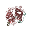
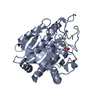
| Structure viewer | Molecule: MolmilJmol/JSmol |
|---|
- Downloads & links
-Download
| PDBx/mmCIF format | 3u3k.cif.gz | 252.4 KB | Display | PDBx/mmCIF format |
|---|---|---|---|---|
| PDB format | pdb3u3k.ent.gz | 203.4 KB | Display | PDB format |
| PDBx/mmJSON format | 3u3k.json.gz | Tree view | PDBx/mmJSON format | |
| Others |  Other downloads Other downloads |
-Validation report
| Arichive directory | https://data.pdbj.org/pub/pdb/validation_reports/u3/3u3kftp://data.pdbj.org/pub/pdb/validation_reports/u3/3u3k | HTTPS FTP |
|---|
-Related structure data
| Related structure data |  3u3jC 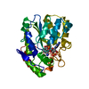 3u3mC 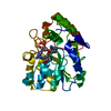 3u3oC 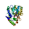 3u3rC 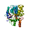 1ls6S C: citing same article ( S: Starting model for refinement |
|---|---|
| Similar structure data |

-Links
 PDBj
PDBj
- Assembly
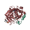
| Deposited unit | 
| ||||||||
|---|---|---|---|---|---|---|---|---|---|
| 1 | 
| ||||||||
| 2 | 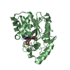
| ||||||||
| Unit cell |
|
-Components
| #1: Protein | Mass: 36391.664 Da / Num. of mol.: 2 Source method: isolated from a genetically manipulated source Source: (gene. exp.) Homo sapiens (human) / Strain: Homo sapiensHuman / Gene: hSULT1A1, OK/SW-cl.88, STP, STP1, SULT1A1 / Plasmid: pET32tr / Production host:  Escherichia coli (E. coli) / Strain (production host): BL21(DE3) / References: UniProt: P50225, aryl sulfotransferase Escherichia coli (E. coli) / Strain (production host): BL21(DE3) / References: UniProt: P50225, aryl sulfotransferase#2: Chemical | Adenosine 3',5'-bisphosphate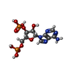  Type: RNA linking / Mass: 427.201 Da / Num. of mol.: 2 / Source method: obtained synthetically / Formula: C10H15N5O10P2 Type: RNA linking / Mass: 427.201 Da / Num. of mol.: 2 / Source method: obtained synthetically / Formula: C10H15N5O10P2#3: Chemical | 2-Naphthol  Mass: 144.170 Da / Num. of mol.: 2 / Source method: obtained synthetically / Formula: C10H8O Mass: 144.170 Da / Num. of mol.: 2 / Source method: obtained synthetically / Formula: C10H8O#4: Water | ChemComp-HOH / | Water Mass: 18.015 Da / Num. of mol.: 113 / Source method: isolated from a natural source / Formula: H2O Mass: 18.015 Da / Num. of mol.: 113 / Source method: isolated from a natural source / Formula: H2OSequence details | THESE CONFLICTS ARISE FROM POLYMORPHISM OF THE HSULT1A1 GENE (H213). THE CLOSEST SEQUENCE TO THE ...THESE CONFLICTS ARISE FROM POLYMORPHI | |
|---|
-Experimental details
-Experiment
| Experiment | Method: X-RAY DIFFRACTION / Number of used crystals: 1 |
|---|
- Sample preparation
| Crystal | Density Matthews: 2.45 Å3/Da / Density % sol: 49.86 % |
|---|---|
| Crystal grow | Temperature: 293 K / Method: vapor diffusion / pH: 8.5 Details: Tris, Polyethylene glycol 3.350, pH 8.5, VAPOR DIFFUSION, temperature 293K |
-Data collection
| Diffraction | Mean temperature: 100 K | |||||||||||||||||||||||||||||||||||||||||||||||||||||||||||||||||||||||||||||||||||||||||||||||||||||||||||||||||||||||||||||||||||||||||||||||||||
|---|---|---|---|---|---|---|---|---|---|---|---|---|---|---|---|---|---|---|---|---|---|---|---|---|---|---|---|---|---|---|---|---|---|---|---|---|---|---|---|---|---|---|---|---|---|---|---|---|---|---|---|---|---|---|---|---|---|---|---|---|---|---|---|---|---|---|---|---|---|---|---|---|---|---|---|---|---|---|---|---|---|---|---|---|---|---|---|---|---|---|---|---|---|---|---|---|---|---|---|---|---|---|---|---|---|---|---|---|---|---|---|---|---|---|---|---|---|---|---|---|---|---|---|---|---|---|---|---|---|---|---|---|---|---|---|---|---|---|---|---|---|---|---|---|---|---|---|---|
| Diffraction source | Source: ROTATING ANODE / Type: RIGAKU RUH3R / Wavelength: 1.54 Å | |||||||||||||||||||||||||||||||||||||||||||||||||||||||||||||||||||||||||||||||||||||||||||||||||||||||||||||||||||||||||||||||||||||||||||||||||||
| Detector | Type: MAR scanner 345 mm plate / Detector: IMAGE PLATE / Date: Nov 1, 2009 | |||||||||||||||||||||||||||||||||||||||||||||||||||||||||||||||||||||||||||||||||||||||||||||||||||||||||||||||||||||||||||||||||||||||||||||||||||
| Radiation | Monochromator: MIRRORS / Protocol: SINGLE WAVELENGTH / Monochromatic (M) / Laue (L): M / Scattering type: x-ray | |||||||||||||||||||||||||||||||||||||||||||||||||||||||||||||||||||||||||||||||||||||||||||||||||||||||||||||||||||||||||||||||||||||||||||||||||||
| Radiation wavelength | Wavelength: 1.54 Å / Relative weight: 1 | |||||||||||||||||||||||||||||||||||||||||||||||||||||||||||||||||||||||||||||||||||||||||||||||||||||||||||||||||||||||||||||||||||||||||||||||||||
| Reflection | Resolution: 2.36→22 Å / Num. obs: 28872 / % possible obs: 94.8 % / Redundancy: 5.5 % / Rmerge(I) obs: 0.089 / Χ2: 1.855 / Net I/σ(I): 11.1 | |||||||||||||||||||||||||||||||||||||||||||||||||||||||||||||||||||||||||||||||||||||||||||||||||||||||||||||||||||||||||||||||||||||||||||||||||||
| Reflection shell |
|
- Processing
| Software |
| |||||||||||||||||||||||||||||||||||||||||||||||||||||||||||||||||||||||||||||||||||||||||||||||||||||||||||||||||||||||||||||||||||||||||||||||||||||||||||||||||||||||||||||||||||||||||||||||||||||||||||||||||||||||||||||||||||||||||||||||||||||||||||||||||||||||||||||||||||||||||||||||||||||||||||||||||||||||||||||||||||||
|---|---|---|---|---|---|---|---|---|---|---|---|---|---|---|---|---|---|---|---|---|---|---|---|---|---|---|---|---|---|---|---|---|---|---|---|---|---|---|---|---|---|---|---|---|---|---|---|---|---|---|---|---|---|---|---|---|---|---|---|---|---|---|---|---|---|---|---|---|---|---|---|---|---|---|---|---|---|---|---|---|---|---|---|---|---|---|---|---|---|---|---|---|---|---|---|---|---|---|---|---|---|---|---|---|---|---|---|---|---|---|---|---|---|---|---|---|---|---|---|---|---|---|---|---|---|---|---|---|---|---|---|---|---|---|---|---|---|---|---|---|---|---|---|---|---|---|---|---|---|---|---|---|---|---|---|---|---|---|---|---|---|---|---|---|---|---|---|---|---|---|---|---|---|---|---|---|---|---|---|---|---|---|---|---|---|---|---|---|---|---|---|---|---|---|---|---|---|---|---|---|---|---|---|---|---|---|---|---|---|---|---|---|---|---|---|---|---|---|---|---|---|---|---|---|---|---|---|---|---|---|---|---|---|---|---|---|---|---|---|---|---|---|---|---|---|---|---|---|---|---|---|---|---|---|---|---|---|---|---|---|---|---|---|---|---|---|---|---|---|---|---|---|---|---|---|---|---|---|---|---|---|---|---|---|---|---|---|---|---|---|---|---|---|---|---|---|---|---|---|---|---|---|---|---|---|---|---|---|---|---|---|---|---|---|---|---|---|---|---|---|---|---|---|---|---|---|
| Refinement | Method to determine structure: MOLECULAR REPLACEMENT Starting model: pdb entry 1LS6 Resolution: 2.36→22 Å / Cor.coef. Fo:Fc: 0.954 / Cor.coef. Fo:Fc free: 0.929 / WRfactor Rfree: 0.2483 / WRfactor Rwork: 0.1991 / Occupancy max: 1 / Occupancy min: 0.17 / FOM work R set: 0.7644 / SU B: 16.593 / SU ML: 0.191 / SU R Cruickshank DPI: 0.4239 / SU Rfree: 0.281 / Cross valid method: THROUGHOUT / σ(F): 0 / ESU R Free: 0.281 / Stereochemistry target values: MAXIMUM LIKELIHOOD Details: HYDROGENS HAVE BEEN ADDED IN THE RIDING POSITIONS U VALUES : WITH TLS ADDED
| |||||||||||||||||||||||||||||||||||||||||||||||||||||||||||||||||||||||||||||||||||||||||||||||||||||||||||||||||||||||||||||||||||||||||||||||||||||||||||||||||||||||||||||||||||||||||||||||||||||||||||||||||||||||||||||||||||||||||||||||||||||||||||||||||||||||||||||||||||||||||||||||||||||||||||||||||||||||||||||||||||||
| Solvent computation | Ion probe radii: 0.8 Å / Shrinkage radii: 0.8 Å / VDW probe radii: 1.4 Å / Solvent model: MASK | |||||||||||||||||||||||||||||||||||||||||||||||||||||||||||||||||||||||||||||||||||||||||||||||||||||||||||||||||||||||||||||||||||||||||||||||||||||||||||||||||||||||||||||||||||||||||||||||||||||||||||||||||||||||||||||||||||||||||||||||||||||||||||||||||||||||||||||||||||||||||||||||||||||||||||||||||||||||||||||||||||||
| Displacement parameters | Biso max: 87.37 Å2 / Biso mean: 44.1194 Å2 / Biso min: 19.93 Å2
| |||||||||||||||||||||||||||||||||||||||||||||||||||||||||||||||||||||||||||||||||||||||||||||||||||||||||||||||||||||||||||||||||||||||||||||||||||||||||||||||||||||||||||||||||||||||||||||||||||||||||||||||||||||||||||||||||||||||||||||||||||||||||||||||||||||||||||||||||||||||||||||||||||||||||||||||||||||||||||||||||||||
| Refinement step | Cycle: LAST / Resolution: 2.36→22 Å
| |||||||||||||||||||||||||||||||||||||||||||||||||||||||||||||||||||||||||||||||||||||||||||||||||||||||||||||||||||||||||||||||||||||||||||||||||||||||||||||||||||||||||||||||||||||||||||||||||||||||||||||||||||||||||||||||||||||||||||||||||||||||||||||||||||||||||||||||||||||||||||||||||||||||||||||||||||||||||||||||||||||
| Refine LS restraints |
| |||||||||||||||||||||||||||||||||||||||||||||||||||||||||||||||||||||||||||||||||||||||||||||||||||||||||||||||||||||||||||||||||||||||||||||||||||||||||||||||||||||||||||||||||||||||||||||||||||||||||||||||||||||||||||||||||||||||||||||||||||||||||||||||||||||||||||||||||||||||||||||||||||||||||||||||||||||||||||||||||||||
| LS refinement shell | Resolution: 2.361→2.422 Å / Total num. of bins used: 20
| |||||||||||||||||||||||||||||||||||||||||||||||||||||||||||||||||||||||||||||||||||||||||||||||||||||||||||||||||||||||||||||||||||||||||||||||||||||||||||||||||||||||||||||||||||||||||||||||||||||||||||||||||||||||||||||||||||||||||||||||||||||||||||||||||||||||||||||||||||||||||||||||||||||||||||||||||||||||||||||||||||||
| Refinement TLS params. | Method: refined / Refine-ID: X-RAY DIFFRACTION
| |||||||||||||||||||||||||||||||||||||||||||||||||||||||||||||||||||||||||||||||||||||||||||||||||||||||||||||||||||||||||||||||||||||||||||||||||||||||||||||||||||||||||||||||||||||||||||||||||||||||||||||||||||||||||||||||||||||||||||||||||||||||||||||||||||||||||||||||||||||||||||||||||||||||||||||||||||||||||||||||||||||
| Refinement TLS group |
|
