 Movie
Movie Controller

[English] 日本語
 Yorodumi
- PDB-3thd: Crystal structure of human beta-galactosidase in complex with 1-d... -
+ Open data

- Basic information
| Entry | Database: PDB / ID: 3thd | ||||||
|---|---|---|---|---|---|---|---|
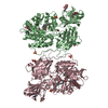
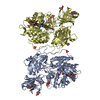
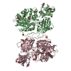
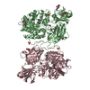
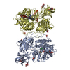
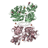
| Title | Crystal structure of human beta-galactosidase in complex with 1-deoxygalactonojirimycin | ||||||
 Components Components | Beta-galactosidase | ||||||
 Keywords Keywords | HYDROLASE / BETA-GALACTOSIDASE / TIM-BARREL DOMAIN / GLYCOSYL HYDROLASE / Glycosylation | ||||||
| Function / homology |  Function and homology information Function and homology informationMPS IV - Morquio syndrome B / response to cortisone / keratan sulfate catabolic process / response to Thyroglobulin triiodothyronine / Defective NEU1 causes sialidosis / Keratan sulfate degradation / galactose catabolic process / heparan sulfate proteoglycan catabolic process / Sialic acid metabolism / HS-GAG degradation ...MPS IV - Morquio syndrome B / response to cortisone / keratan sulfate catabolic process / response to Thyroglobulin triiodothyronine / Defective NEU1 causes sialidosis / Keratan sulfate degradation / galactose catabolic process / heparan sulfate proteoglycan catabolic process / Sialic acid metabolism / HS-GAG degradation / glycosphingolipid catabolic process / galactoside binding / Glycosphingolipid catabolism / beta-galactosidase / vacuole / beta-galactosidase activity / lysosomal lumen / azurophil granule lumen / ficolin-1-rich granule lumen / carbohydrate metabolic process / intracellular membrane-bounded organelle / Neutrophil degranulation / perinuclear region of cytoplasm / Golgi apparatus / protein homodimerization activity / extracellular exosome / extracellular region / cytoplasmSimilarity search - Function | ||||||
| Biological species |  Homo sapiens (human) Homo sapiens (human) | ||||||
| Method | X-RAY DIFFRACTION / SYNCHROTRON / MOLECULAR REPLACEMENT / Resolution: 1.79 Å | ||||||
 Authors Authors | Ohto, U. / Shimizu, T. | ||||||
 Citation Citation | Journal: J.Biol.Chem. / Year: 2012 Title: Crystal structure of human beta-galactosidase: structural basis of Gm1 gangliosidosis and morquio B diseases Authors: Ohto, U. / Usui, K. / Ochi, T. / Yuki, K. / Satow, Y. / Shimizu, T. | ||||||
| History |
|
- Structure visualization
| Structure viewer | Molecule: MolmilJmol/JSmol |
|---|
- Downloads & links
-Download
| PDBx/mmCIF format | 3thd.cif.gz | 983.2 KB | Display | PDBx/mmCIF format |
|---|---|---|---|---|
| PDB format | pdb3thd.ent.gz | 817.2 KB | Display | PDB format |
| PDBx/mmJSON format | 3thd.json.gz | Tree view | PDBx/mmJSON format | |
| Others |  Other downloads Other downloads |
-Validation report
| Arichive directory | https://data.pdbj.org/pub/pdb/validation_reports/th/3thdftp://data.pdbj.org/pub/pdb/validation_reports/th/3thd | HTTPS FTP |
|---|
-Related structure data
| Related structure data | 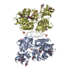 3thcC  3d3aS C: citing same article ( S: Starting model for refinement |
|---|---|
| Similar structure data |


-Links
 PDBj
PDBj

- Assembly
| Deposited unit | 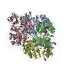
| ||||||||
|---|---|---|---|---|---|---|---|---|---|
| 1 | 
| ||||||||
| 2 | 
| ||||||||
| Unit cell |
|
-Components
-Protein / Sugars , 2 types, 20 molecules ABCD
| #1: Protein | Mass: 73665.336 Da / Num. of mol.: 4 Source method: isolated from a genetically manipulated source Source: (gene. exp.) Homo sapiens (human) / Gene: GLB1, ELNR1 / Production host:  Pichia pastoris (fungus) / References: UniProt: P16278, beta-galactosidase Pichia pastoris (fungus) / References: UniProt: P16278, beta-galactosidase#2: Sugar | ChemComp-NAG / N-Acetylglucosamine Type: D-saccharide, beta linking / Mass: 221.208 Da / Num. of mol.: 16 Type: D-saccharide, beta linking / Mass: 221.208 Da / Num. of mol.: 16Source method: isolated from a genetically manipulated source Formula: C8H15NO6 |
|---|
-Non-polymers , 5 types, 2208 molecules 


| #3: Chemical | ChemComp-CL / Chloride Mass: 35.453 Da / Num. of mol.: 4 / Source method: obtained synthetically / Formula: Cl Mass: 35.453 Da / Num. of mol.: 4 / Source method: obtained synthetically / Formula: Cl#4: Chemical | ChemComp-DGJ / ( Migalastat Mass: 163.172 Da / Num. of mol.: 4 / Source method: obtained synthetically / Formula: C6H13NO4 / Comment: medication, Galafold*YM Mass: 163.172 Da / Num. of mol.: 4 / Source method: obtained synthetically / Formula: C6H13NO4 / Comment: medication, Galafold*YM#5: Chemical | ChemComp-SO4 / Sulfate Mass: 96.063 Da / Num. of mol.: 8 / Source method: obtained synthetically / Formula: SO4 Mass: 96.063 Da / Num. of mol.: 8 / Source method: obtained synthetically / Formula: SO4#6: Chemical | ChemComp-EDO / Ethylene glycol Mass: 62.068 Da / Num. of mol.: 8 / Source method: obtained synthetically / Formula: C2H6O2 Mass: 62.068 Da / Num. of mol.: 8 / Source method: obtained synthetically / Formula: C2H6O2#7: Water | ChemComp-HOH / | WaterMass: 18.015 Da / Num. of mol.: 2184 / Source method: isolated from a natural source / Formula: H2O |
|---|
-Experimental details
-Experiment
| Experiment | Method: X-RAY DIFFRACTION / Number of used crystals: 1 |
|---|
- Sample preparation
| Crystal | Density Matthews: 2.62 Å3/Da / Density % sol: 52.99 % |
|---|---|
| Crystal grow | Temperature: 293 K / Method: vapor diffusion, sitting drop / pH: 8 Details: 20% (w/v) PEG3350 0.2M ammonium sulfate 100mM Tris HCl pH 8.0 , VAPOR DIFFUSION, SITTING DROP, temperature 293K |
-Data collection
| Diffraction | Mean temperature: 95 K |
|---|---|
| Diffraction source | Source: SYNCHROTRON / Site: Photon Factory  / Beamline: AR-NW12A / Wavelength: 1 Å / Beamline: AR-NW12A / Wavelength: 1 Å |
| Detector | Type: ADSC QUANTUM 270 / Detector: CCD / Date: Oct 24, 2009 |
| Radiation | Protocol: SINGLE WAVELENGTH / Monochromatic (M) / Laue (L): M / Scattering type: x-ray |
| Radiation wavelength | Wavelength: 1 Å / Relative weight: 1 |
| Reflection | Resolution: 1.79→29.29 Å / Num. obs: 249160 |
- Processing
| Software |
| |||||||||||||||||||||||||||||||||||||||||||||||||||||||||||||||||||||||||||||||||||||||||||||||||||||||||||||||||||||||||||||||||||||||||||||||||||||||||||||||||||||||||||||||||||||||||||||||||||||||||||||||||||||||||||||||||
|---|---|---|---|---|---|---|---|---|---|---|---|---|---|---|---|---|---|---|---|---|---|---|---|---|---|---|---|---|---|---|---|---|---|---|---|---|---|---|---|---|---|---|---|---|---|---|---|---|---|---|---|---|---|---|---|---|---|---|---|---|---|---|---|---|---|---|---|---|---|---|---|---|---|---|---|---|---|---|---|---|---|---|---|---|---|---|---|---|---|---|---|---|---|---|---|---|---|---|---|---|---|---|---|---|---|---|---|---|---|---|---|---|---|---|---|---|---|---|---|---|---|---|---|---|---|---|---|---|---|---|---|---|---|---|---|---|---|---|---|---|---|---|---|---|---|---|---|---|---|---|---|---|---|---|---|---|---|---|---|---|---|---|---|---|---|---|---|---|---|---|---|---|---|---|---|---|---|---|---|---|---|---|---|---|---|---|---|---|---|---|---|---|---|---|---|---|---|---|---|---|---|---|---|---|---|---|---|---|---|---|---|---|---|---|---|---|---|---|---|---|---|---|---|---|---|---|
| Refinement | Method to determine structure: MOLECULAR REPLACEMENT Starting model: 3D3A Resolution: 1.79→29.29 Å / Cor.coef. Fo:Fc: 0.933 / Cor.coef. Fo:Fc free: 0.914 / SU B: 5.008 / SU ML: 0.087 / Cross valid method: THROUGHOUT / ESU R Free: 0.127 / Stereochemistry target values: MAXIMUM LIKELIHOOD / Details: HYDROGENS HAVE BEEN USED IF PRESENT IN THE INPUT
| |||||||||||||||||||||||||||||||||||||||||||||||||||||||||||||||||||||||||||||||||||||||||||||||||||||||||||||||||||||||||||||||||||||||||||||||||||||||||||||||||||||||||||||||||||||||||||||||||||||||||||||||||||||||||||||||||
| Solvent computation | Ion probe radii: 0.8 Å / Shrinkage radii: 0.8 Å / VDW probe radii: 1.2 Å / Solvent model: BABINET MODEL WITH MASK | |||||||||||||||||||||||||||||||||||||||||||||||||||||||||||||||||||||||||||||||||||||||||||||||||||||||||||||||||||||||||||||||||||||||||||||||||||||||||||||||||||||||||||||||||||||||||||||||||||||||||||||||||||||||||||||||||
| Displacement parameters | Biso mean: 20.053 Å2
| |||||||||||||||||||||||||||||||||||||||||||||||||||||||||||||||||||||||||||||||||||||||||||||||||||||||||||||||||||||||||||||||||||||||||||||||||||||||||||||||||||||||||||||||||||||||||||||||||||||||||||||||||||||||||||||||||
| Refinement step | Cycle: LAST / Resolution: 1.79→29.29 Å
| |||||||||||||||||||||||||||||||||||||||||||||||||||||||||||||||||||||||||||||||||||||||||||||||||||||||||||||||||||||||||||||||||||||||||||||||||||||||||||||||||||||||||||||||||||||||||||||||||||||||||||||||||||||||||||||||||
| Refine LS restraints |
| |||||||||||||||||||||||||||||||||||||||||||||||||||||||||||||||||||||||||||||||||||||||||||||||||||||||||||||||||||||||||||||||||||||||||||||||||||||||||||||||||||||||||||||||||||||||||||||||||||||||||||||||||||||||||||||||||
| LS refinement shell | Resolution: 1.794→1.841 Å / Total num. of bins used: 20
| |||||||||||||||||||||||||||||||||||||||||||||||||||||||||||||||||||||||||||||||||||||||||||||||||||||||||||||||||||||||||||||||||||||||||||||||||||||||||||||||||||||||||||||||||||||||||||||||||||||||||||||||||||||||||||||||||
| Refinement TLS params. | Method: refined / Refine-ID: X-RAY DIFFRACTION
| |||||||||||||||||||||||||||||||||||||||||||||||||||||||||||||||||||||||||||||||||||||||||||||||||||||||||||||||||||||||||||||||||||||||||||||||||||||||||||||||||||||||||||||||||||||||||||||||||||||||||||||||||||||||||||||||||
| Refinement TLS group |
|
