 Movie
Movie Controller

[English] 日本語
 Yorodumi
- PDB-3wf3: Crystal structure of human beta-galactosidase mutant I51T in comp... -
+ Open data

- Basic information
| Entry | Database: PDB / ID: 3wf3 | ||||||
|---|---|---|---|---|---|---|---|
| Title | Crystal structure of human beta-galactosidase mutant I51T in complex with Galactose | ||||||
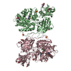
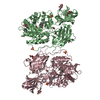
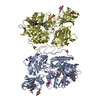
 Components Components | Beta-galactosidase | ||||||
 Keywords Keywords | HYDROLASE / GLYCOSYL HYDROLASE / TIM-BARREL DOMAIN | ||||||
| Function / homology |  Function and homology information Function and homology informationMPS IV - Morquio syndrome B / response to cortisone / keratan sulfate catabolic process / response to Thyroglobulin triiodothyronine / Defective NEU1 causes sialidosis / Keratan sulfate degradation / galactose catabolic process / heparan sulfate proteoglycan catabolic process / Sialic acid metabolism / HS-GAG degradation ...MPS IV - Morquio syndrome B / response to cortisone / keratan sulfate catabolic process / response to Thyroglobulin triiodothyronine / Defective NEU1 causes sialidosis / Keratan sulfate degradation / galactose catabolic process / heparan sulfate proteoglycan catabolic process / Sialic acid metabolism / HS-GAG degradation / galactoside binding / glycosphingolipid catabolic process / Glycosphingolipid catabolism / beta-galactosidase / vacuole / beta-galactosidase activity / lysosomal lumen / azurophil granule lumen / ficolin-1-rich granule lumen / carbohydrate metabolic process / intracellular membrane-bounded organelle / Neutrophil degranulation / perinuclear region of cytoplasm / Golgi apparatus / protein homodimerization activity / extracellular exosome / extracellular region / cytoplasmSimilarity search - Function | ||||||
| Biological species |  Homo sapiens (human) Homo sapiens (human) | ||||||
| Method | X-RAY DIFFRACTION / SYNCHROTRON / MOLECULAR REPLACEMENT / Resolution: 2.15 Å | ||||||
 Authors Authors | Suzuki, H. / Ohto, U. / Shimizu, T. | ||||||
 Citation Citation | Journal: to be published Title: Structural basis of pharmacological chaperoning for human beta-galactosidase Authors: Suzuki, H. / Ohto, U. / Higaki, K. / Mena-Barragan, T. / Aguilar-Moncayo, M. / Ortiz Mellet, C. / Nanba, E. / Garcia Fernandez, J.M. / Suzuki, Y. / Shimizu, T. | ||||||
| History |
|
- Structure visualization
| Structure viewer | Molecule: MolmilJmol/JSmol |
|---|
- Downloads & links
-Download
| PDBx/mmCIF format | 3wf3.cif.gz | 517.4 KB | Display | PDBx/mmCIF format |
|---|---|---|---|---|
| PDB format | pdb3wf3.ent.gz | 422.2 KB | Display | PDB format |
| PDBx/mmJSON format | 3wf3.json.gz | Tree view | PDBx/mmJSON format | |
| Others |  Other downloads Other downloads |
-Validation report
| Arichive directory | https://data.pdbj.org/pub/pdb/validation_reports/wf/3wf3ftp://data.pdbj.org/pub/pdb/validation_reports/wf/3wf3 | HTTPS FTP |
|---|
-Related structure data
| Related structure data | 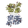 3wezC 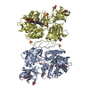 3wf0C 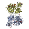 3wf1C  3wf2C 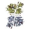 3wf4C 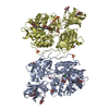 3thcS C: citing same article ( S: Starting model for refinement |
|---|---|
| Similar structure data |


-Links
 PDBj
PDBj


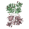
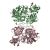
- Assembly
| Deposited unit | 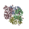
| ||||||||
|---|---|---|---|---|---|---|---|---|---|
| 1 | 
| ||||||||
| 2 | 
| ||||||||
| Unit cell |
|
-Components
-Protein , 1 types, 4 molecules ABCD
| #1: Protein | / Acid beta-galactosidase / Lactase / Elastin receptor 1 Mass: 76606.297 Da / Num. of mol.: 4 / Mutation: I51T Source method: isolated from a genetically manipulated source Source: (gene. exp.) Homo sapiens (human) / Gene: ELNR1, GLB1 / Production host:  Pichia pastoris (fungus) / References: UniProt: P16278, beta-galactosidase Pichia pastoris (fungus) / References: UniProt: P16278, beta-galactosidase |
|---|
-Sugars , 2 types, 20 molecules 

| #2: Sugar | ChemComp-NAG / N-Acetylglucosamine Type: D-saccharide, beta linking / Mass: 221.208 Da / Num. of mol.: 16 Type: D-saccharide, beta linking / Mass: 221.208 Da / Num. of mol.: 16Source method: isolated from a genetically manipulated source Formula: C8H15NO6 #3: Sugar | ChemComp-GAL / Galactose Type: D-saccharide, beta linking / Mass: 180.156 Da / Num. of mol.: 4 Type: D-saccharide, beta linking / Mass: 180.156 Da / Num. of mol.: 4Source method: isolated from a genetically manipulated source Formula: C6H12O6 |
|---|
-Non-polymers , 4 types, 1448 molecules 


| #4: Chemical | ChemComp-CL / Chloride Mass: 35.453 Da / Num. of mol.: 4 / Source method: obtained synthetically / Formula: Cl Mass: 35.453 Da / Num. of mol.: 4 / Source method: obtained synthetically / Formula: Cl#5: Chemical | ChemComp-SO4 / Sulfate Mass: 96.063 Da / Num. of mol.: 8 / Source method: obtained synthetically / Formula: SO4 Mass: 96.063 Da / Num. of mol.: 8 / Source method: obtained synthetically / Formula: SO4#6: Chemical | ChemComp-EDO / Ethylene glycol Mass: 62.068 Da / Num. of mol.: 8 / Source method: obtained synthetically / Formula: C2H6O2 Mass: 62.068 Da / Num. of mol.: 8 / Source method: obtained synthetically / Formula: C2H6O2#7: Water | ChemComp-HOH / | WaterMass: 18.015 Da / Num. of mol.: 1428 / Source method: isolated from a natural source / Formula: H2O |
|---|
-Experimental details
-Experiment
| Experiment | Method: X-RAY DIFFRACTION / Number of used crystals: 1 |
|---|
- Sample preparation
| Crystal | Density Matthews: 2.62 Å3/Da / Density % sol: 53.02 % |
|---|---|
| Crystal grow | Temperature: 277 K / Method: vapor diffusion, sitting drop / pH: 7.5 Details: PEG 3350, Ammonium sulfate, HEPES, pH 7.5, vapor diffusion, sittingdrop, temperature 277K |
-Data collection
| Diffraction | Mean temperature: 95 K |
|---|---|
| Diffraction source | Source: SYNCHROTRON / Site: Photon Factory  / Beamline: AR-NE3A / Wavelength: 1 Å / Beamline: AR-NE3A / Wavelength: 1 Å |
| Detector | Type: ADSC QUANTUM 270 / Detector: CCD / Date: Jan 20, 2013 |
| Radiation | Protocol: SINGLE WAVELENGTH / Monochromatic (M) / Laue (L): M / Scattering type: x-ray |
| Radiation wavelength | Wavelength: 1 Å / Relative weight: 1 |
| Reflection | Resolution: 2.15→50 Å / Num. obs: 160267 / % possible obs: 96.4 % / Redundancy: 3.2 % / Rmerge(I) obs: 0.154 / Net I/σ(I): 15.5 |
| Reflection shell | Resolution: 2.15→2.19 Å / Redundancy: 3.3 % / Rmerge(I) obs: 0.478 / Mean I/σ(I) obs: 3.5 / % possible all: 98.6 |
- Processing
| Software |
| ||||||||||||||||||||||||||||||||||||||||||||||||||||||||||||
|---|---|---|---|---|---|---|---|---|---|---|---|---|---|---|---|---|---|---|---|---|---|---|---|---|---|---|---|---|---|---|---|---|---|---|---|---|---|---|---|---|---|---|---|---|---|---|---|---|---|---|---|---|---|---|---|---|---|---|---|---|---|
| Refinement | Method to determine structure: MOLECULAR REPLACEMENT Starting model: 3THC Resolution: 2.15→27.3 Å / Cor.coef. Fo:Fc: 0.941 / Cor.coef. Fo:Fc free: 0.913 / Occupancy max: 1 / Occupancy min: 0.5 / SU B: 5.538 / SU ML: 0.144 / Cross valid method: THROUGHOUT / σ(F): 0 / ESU R: 0.243 / ESU R Free: 0.2 / Stereochemistry target values: MAXIMUM LIKELIHOOD Details: HYDROGENS HAVE BEEN ADDED IN THE RIDING POSITIONS U VALUES: REFINED INDIVIDUALLY
| ||||||||||||||||||||||||||||||||||||||||||||||||||||||||||||
| Solvent computation | Ion probe radii: 0.8 Å / Shrinkage radii: 0.8 Å / VDW probe radii: 1.2 Å / Solvent model: MASK | ||||||||||||||||||||||||||||||||||||||||||||||||||||||||||||
| Displacement parameters | Biso max: 103.95 Å2 / Biso mean: 29.4081 Å2 / Biso min: 13.66 Å2
| ||||||||||||||||||||||||||||||||||||||||||||||||||||||||||||
| Refinement step | Cycle: LAST / Resolution: 2.15→27.3 Å
| ||||||||||||||||||||||||||||||||||||||||||||||||||||||||||||
| Refine LS restraints |
| ||||||||||||||||||||||||||||||||||||||||||||||||||||||||||||
| LS refinement shell | Resolution: 2.15→2.205 Å / Total num. of bins used: 20
|
