 Movie
Movie Controller

[English] 日本語
 Yorodumi
- PDB-3te9: 1.8 Angstrom Resolution Crystal Structure of K135M Mutant of Tran... -
+ Open data

- Basic information
| Entry | Database: PDB / ID: 3te9 | ||||||
|---|---|---|---|---|---|---|---|
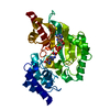
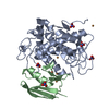
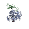
| Title | 1.8 Angstrom Resolution Crystal Structure of K135M Mutant of Transaldolase B (TalA) from Francisella tularensis in Complex with Fructose 6-phosphate | ||||||
 Components Components | Transaldolase | ||||||
 Keywords Keywords | TRANSFERASE / Structural Genomics / Center for Structural Genomics of Infectious Diseases / CSGID / Alpha-Beta Barrel/TIM Barrel / sedoheptulose-7-phosphate:D-glyceraldehyde-3-phosphate glyceronetransferase activity | ||||||
| Function / homology |  Function and homology informationtransaldolase / transaldolase activity / pentose-phosphate shunt, non-oxidative branch / carbohydrate metabolic process / cytosol Function and homology informationtransaldolase / transaldolase activity / pentose-phosphate shunt, non-oxidative branch / carbohydrate metabolic process / cytosolSimilarity search - Function | ||||||
| Biological species |  Francisella tularensis subsp. tularensis (bacteria) Francisella tularensis subsp. tularensis (bacteria) | ||||||
| Method | X-RAY DIFFRACTION / SYNCHROTRON / MOLECULAR REPLACEMENT / Resolution: 1.8 Å | ||||||
 Authors Authors | Minasov, G. / Light, S.H. / Halavaty, A. / Shuvalova, L. / Papazisi, L. / Anderson, W.F. / Center for Structural Genomics of Infectious Diseases (CSGID) | ||||||
 Citation Citation | Journal: Acta Crystallogr.,Sect.D / Year: 2014 Title: Adherence to Burgi-Dunitz stereochemical principles requires significant structural rearrangements in Schiff-base formation: insights from transaldolase complexes. Authors: Light, S.H. / Minasov, G. / Duban, M.E. / Anderson, W.F. | ||||||
| History |
|
- Structure visualization
| Structure viewer | Molecule: MolmilJmol/JSmol |
|---|
- Downloads & links
-Download
| PDBx/mmCIF format | 3te9.cif.gz | 285 KB | Display | PDBx/mmCIF format |
|---|---|---|---|---|
| PDB format | pdb3te9.ent.gz | 231.8 KB | Display | PDB format |
| PDBx/mmJSON format | 3te9.json.gz | Tree view | PDBx/mmJSON format | |
| Others |  Other downloads Other downloads |
-Validation report
| Arichive directory | https://data.pdbj.org/pub/pdb/validation_reports/te/3te9ftp://data.pdbj.org/pub/pdb/validation_reports/te/3te9 | HTTPS FTP |
|---|
-Related structure data
| Related structure data |  3tk7C 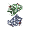 3tkfC  3tnoC  4e0cC  3igxS S: Starting model for refinement C: citing same article ( |
|---|---|
| Similar structure data | |
| Other databases |

-Links
 PDBj
PDBj- Assembly
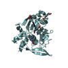
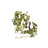
| Deposited unit | 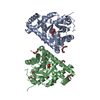
| ||||||||
|---|---|---|---|---|---|---|---|---|---|
| 1 | 
| ||||||||
| 2 | 
| ||||||||
| 3 |
| ||||||||
| Unit cell |
|
-Components
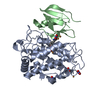
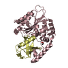
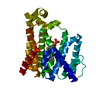
-Protein , 1 types, 2 molecules AB
| #1: Protein | Mass: 38478.090 Da / Num. of mol.: 2 / Mutation: K135M Source method: isolated from a genetically manipulated source Source: (gene. exp.) Francisella tularensis subsp. tularensis (bacteria)Strain: SCHU S4 / Gene: FTT_1093c, talA / Plasmid: pMCSG7 / Production host: Escherichia coli (E. coli) / Strain (production host): BL21 magic / References: UniProt: Q5NFX0, transaldolase |
|---|
-Non-polymers , 5 types, 620 molecules 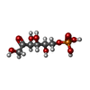


| #2: Chemical | ChemComp-F6R / Fructose 6-phosphate Mass: 260.136 Da / Num. of mol.: 1 / Source method: obtained synthetically / Formula: C6H13O9P Mass: 260.136 Da / Num. of mol.: 1 / Source method: obtained synthetically / Formula: C6H13O9P | ||||||
|---|---|---|---|---|---|---|---|
| #3: Chemical | Diethylene glycol Mass: 106.120 Da / Num. of mol.: 2 / Source method: obtained synthetically / Formula: C4H10O3 Mass: 106.120 Da / Num. of mol.: 2 / Source method: obtained synthetically / Formula: C4H10O3#4: Chemical | ChemComp-PGE / Polyethylene glycol Mass: 150.173 Da / Num. of mol.: 6 / Source method: obtained synthetically / Formula: C6H14O4 Mass: 150.173 Da / Num. of mol.: 6 / Source method: obtained synthetically / Formula: C6H14O4#5: Chemical | ChemComp-PO4 / | Phosphate Mass: 94.971 Da / Num. of mol.: 1 / Source method: obtained synthetically / Formula: PO4 Mass: 94.971 Da / Num. of mol.: 1 / Source method: obtained synthetically / Formula: PO4#6: Water | ChemComp-HOH / | WaterMass: 18.015 Da / Num. of mol.: 610 / Source method: isolated from a natural source / Formula: H2O |
-Experimental details
-Experiment
| Experiment | Method: X-RAY DIFFRACTION / Number of used crystals: 1 |
|---|
- Sample preparation
| Crystal | Density Matthews: 2.17 Å3/Da / Density % sol: 43.34 % |
|---|---|
| Crystal grow | Temperature: 295 K / Method: vapor diffusion, sitting drop / pH: 8.3 Details: Protein: 9.4mG/mL, 0.5M Sodium chloride, 0.1M TRIS-HCl, 0.02M Fructose 6-phosphate; Screen: PEG's (H1), 0.2M Potassium/Sodium tartrate, 20% (w/v) PEG3350., pH 8.3, VAPOR DIFFUSION, SITTING DROP, temperature 295K |
-Data collection
| Diffraction | Mean temperature: 100 K |
|---|---|
| Diffraction source | Source: SYNCHROTRON / Site: APS  / Beamline: 21-ID-F / Wavelength: 0.97872 Å / Beamline: 21-ID-F / Wavelength: 0.97872 Å |
| Detector | Type: MARMOSAIC 225 mm CCD / Detector: CCD / Date: Jul 28, 2011 / Details: Beryllium lenses |
| Radiation | Monochromator: Diamond / Protocol: SINGLE WAVELENGTH / Monochromatic (M) / Laue (L): M / Scattering type: x-ray |
| Radiation wavelength | Wavelength: 0.97872 Å / Relative weight: 1 |
| Reflection | Resolution: 1.8→30 Å / Num. all: 63010 / Num. obs: 63010 / % possible obs: 100 % / Observed criterion σ(I): -3 / Redundancy: 7.2 % / Biso Wilson estimate: 24.3 Å2 / Rmerge(I) obs: 0.075 / Net I/σ(I): 24.5 |
| Reflection shell | Resolution: 1.8→1.83 Å / Redundancy: 7.2 % / Rmerge(I) obs: 0.582 / Mean I/σ(I) obs: 3.7 / Num. unique all: 3096 / % possible all: 100 |
- Processing
| Software |
| |||||||||||||||||||||||||||||||||||||||||||||||||||||||||||||||||||||||||||||||||||||||||||||||||||||||||||||||||||||||||||||||||||||||||||||||||||||||||||||||||||||||||||||||||||||||||||||||||||||||||||||||||||||||||||||||||||||||||||||||||||||||||||||||||||||||||||||||||||
|---|---|---|---|---|---|---|---|---|---|---|---|---|---|---|---|---|---|---|---|---|---|---|---|---|---|---|---|---|---|---|---|---|---|---|---|---|---|---|---|---|---|---|---|---|---|---|---|---|---|---|---|---|---|---|---|---|---|---|---|---|---|---|---|---|---|---|---|---|---|---|---|---|---|---|---|---|---|---|---|---|---|---|---|---|---|---|---|---|---|---|---|---|---|---|---|---|---|---|---|---|---|---|---|---|---|---|---|---|---|---|---|---|---|---|---|---|---|---|---|---|---|---|---|---|---|---|---|---|---|---|---|---|---|---|---|---|---|---|---|---|---|---|---|---|---|---|---|---|---|---|---|---|---|---|---|---|---|---|---|---|---|---|---|---|---|---|---|---|---|---|---|---|---|---|---|---|---|---|---|---|---|---|---|---|---|---|---|---|---|---|---|---|---|---|---|---|---|---|---|---|---|---|---|---|---|---|---|---|---|---|---|---|---|---|---|---|---|---|---|---|---|---|---|---|---|---|---|---|---|---|---|---|---|---|---|---|---|---|---|---|---|---|---|---|---|---|---|---|---|---|---|---|---|---|---|---|---|---|---|---|---|---|---|---|---|---|---|---|---|---|---|---|---|---|---|---|
| Refinement | Method to determine structure: MOLECULAR REPLACEMENT Starting model: 3IGX Resolution: 1.8→29.58 Å / Cor.coef. Fo:Fc: 0.97 / Cor.coef. Fo:Fc free: 0.959 / SU B: 4.729 / SU ML: 0.068 Isotropic thermal model: Thermal Factors Individually Refined Cross valid method: THROUGHOUT / ESU R Free: 0.11 / Stereochemistry target values: MAXIMUM LIKELIHOOD / Details: HYDROGENS HAVE BEEN ADDED IN THE RIDING POSITIONS
| |||||||||||||||||||||||||||||||||||||||||||||||||||||||||||||||||||||||||||||||||||||||||||||||||||||||||||||||||||||||||||||||||||||||||||||||||||||||||||||||||||||||||||||||||||||||||||||||||||||||||||||||||||||||||||||||||||||||||||||||||||||||||||||||||||||||||||||||||||
| Solvent computation | Ion probe radii: 0.8 Å / Shrinkage radii: 0.8 Å / VDW probe radii: 1.2 Å / Solvent model: MASK | |||||||||||||||||||||||||||||||||||||||||||||||||||||||||||||||||||||||||||||||||||||||||||||||||||||||||||||||||||||||||||||||||||||||||||||||||||||||||||||||||||||||||||||||||||||||||||||||||||||||||||||||||||||||||||||||||||||||||||||||||||||||||||||||||||||||||||||||||||
| Displacement parameters | Biso mean: 21.432 Å2
| |||||||||||||||||||||||||||||||||||||||||||||||||||||||||||||||||||||||||||||||||||||||||||||||||||||||||||||||||||||||||||||||||||||||||||||||||||||||||||||||||||||||||||||||||||||||||||||||||||||||||||||||||||||||||||||||||||||||||||||||||||||||||||||||||||||||||||||||||||
| Refinement step | Cycle: LAST / Resolution: 1.8→29.58 Å
| |||||||||||||||||||||||||||||||||||||||||||||||||||||||||||||||||||||||||||||||||||||||||||||||||||||||||||||||||||||||||||||||||||||||||||||||||||||||||||||||||||||||||||||||||||||||||||||||||||||||||||||||||||||||||||||||||||||||||||||||||||||||||||||||||||||||||||||||||||
| Refine LS restraints |
| |||||||||||||||||||||||||||||||||||||||||||||||||||||||||||||||||||||||||||||||||||||||||||||||||||||||||||||||||||||||||||||||||||||||||||||||||||||||||||||||||||||||||||||||||||||||||||||||||||||||||||||||||||||||||||||||||||||||||||||||||||||||||||||||||||||||||||||||||||
| LS refinement shell | Resolution: 1.8→1.847 Å / Total num. of bins used: 20
| |||||||||||||||||||||||||||||||||||||||||||||||||||||||||||||||||||||||||||||||||||||||||||||||||||||||||||||||||||||||||||||||||||||||||||||||||||||||||||||||||||||||||||||||||||||||||||||||||||||||||||||||||||||||||||||||||||||||||||||||||||||||||||||||||||||||||||||||||||
| Refinement TLS params. | Method: refined / Refine-ID: X-RAY DIFFRACTION
| |||||||||||||||||||||||||||||||||||||||||||||||||||||||||||||||||||||||||||||||||||||||||||||||||||||||||||||||||||||||||||||||||||||||||||||||||||||||||||||||||||||||||||||||||||||||||||||||||||||||||||||||||||||||||||||||||||||||||||||||||||||||||||||||||||||||||||||||||||
| Refinement TLS group |
|
