 Movie
Movie Controller

[English] 日本語
 Yorodumi
- PDB-4hcp: crystal structure of Burkholderia pseudomallei effector protein c... -
+ Open data

- Basic information
| Entry | Database: PDB / ID: 4hcp | ||||||
|---|---|---|---|---|---|---|---|
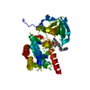
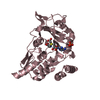
| Title | crystal structure of Burkholderia pseudomallei effector protein chbp in complex with nedd8 | ||||||
 Components Components |
| ||||||
 Keywords Keywords |  PROTEIN BINDING / deamidase / Alpha/Beta/Alpha fold / Deamidation / NEDD8/Ubiquitin / bacterial cytosol PROTEIN BINDING / deamidase / Alpha/Beta/Alpha fold / Deamidation / NEDD8/Ubiquitin / bacterial cytosol | ||||||
| Function / homology |  Function and homology information Function and homology informationsymbiont-mediated perturbation of host cell cycle progression / protein-glutamine glutaminase activity / protein-glutamine glutaminase / regulation of proteolysis / protein neddylation / TGF-beta receptor signaling activates SMADs / anatomical structure morphogenesis / Iron uptake and transport / protein modification process / protein localization ...symbiont-mediated perturbation of host cell cycle progression / protein-glutamine glutaminase activity / protein-glutamine glutaminase / regulation of proteolysis / protein neddylation / TGF-beta receptor signaling activates SMADs / anatomical structure morphogenesis / Iron uptake and transport / protein modification process / protein localization / modification-dependent protein catabolic process / protein tag activity / UCH proteinases / Cargo recognition for clathrin-mediated endocytosis / Neddylation / toxin activity / ubiquitin-dependent protein catabolic process / ubiquitin protein ligase binding / host cell nucleus / regulation of transcription by RNA polymerase II / proteolysis / extracellular exosome / extracellular region / nucleoplasm / nucleus / cytosolSimilarity search - Function | ||||||
| Biological species |  Burkholderia pseudomallei (bacteria) Burkholderia pseudomallei (bacteria) Homo sapiens (human) Homo sapiens (human) | ||||||
| Method | X-RAY DIFFRACTION / SYNCHROTRON / MOLECULAR REPLACEMENT / Resolution: 2.52 Å | ||||||
 Authors Authors | Yao, Q. / Shao, F. | ||||||
 Citation Citation | Journal: Proc.Natl.Acad.Sci.USA / Year: 2012 Title: Structural mechanism of ubiquitin and NEDD8 deamidation catalyzed by bacterial effectors that induce macrophage-specific apoptosis. Authors: Yao, Q. / Cui, J. / Wang, J. / Li, T. / Wan, X. / Luo, T. / Gong, Y.N. / Xu, Y. / Huang, N. / Shao, F. | ||||||
| History |
|
- Structure visualization
| Structure viewer | Molecule: MolmilJmol/JSmol |
|---|
- Downloads & links
-Download
| PDBx/mmCIF format | 4hcp.cif.gz | 77.4 KB | Display | PDBx/mmCIF format |
|---|---|---|---|---|
| PDB format | pdb4hcp.ent.gz | 57.4 KB | Display | PDB format |
| PDBx/mmJSON format | 4hcp.json.gz | Tree view | PDBx/mmJSON format | |
| Others |  Other downloads Other downloads |
-Validation report
| Arichive directory | https://data.pdbj.org/pub/pdb/validation_reports/hc/4hcpftp://data.pdbj.org/pub/pdb/validation_reports/hc/4hcp | HTTPS FTP |
|---|
-Related structure data


-Links
 PDBj
PDBj

- Assembly
| Deposited unit | 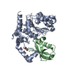
| ||||||||
|---|---|---|---|---|---|---|---|---|---|
| 1 |
| ||||||||
| Unit cell |
|
-Components
| #1: Protein | Mass: 28557.408 Da / Num. of mol.: 1 / Fragment: unp residues 78-382 / Mutation: A156C Source method: isolated from a genetically manipulated source Source: (gene. exp.) Burkholderia pseudomallei (bacteria) / Strain: K96243 / Gene: BPSS1385 / Production host: Escherichia coli (E. coli) / Strain (production host): BL21(DE3) / References: UniProt: Q63KH5 |
|---|---|
| #2: Protein | / Neddylin / Neural precursor cell expressed developmentally down-regulated protein 8 / NEDD-8 / ...Neddylin / Neural precursor cell expressed developmentally down-regulated protein 8 / NEDD-8 / Ubiquitin-like protein Nedd8 Mass: 8716.133 Da / Num. of mol.: 1 / Fragment: unp residues 1-76 Source method: isolated from a genetically manipulated source Source: (gene. exp.) Homo sapiens (human) / Gene: NEDD8 / Production host: Escherichia coli (E. coli) / Strain (production host): BL21(DE3) / References: UniProt: Q15843 |
| #3: Chemical | ChemComp-SO4 / Sulfate  Mass: 96.063 Da / Num. of mol.: 1 / Source method: obtained synthetically / Formula: SO4 Mass: 96.063 Da / Num. of mol.: 1 / Source method: obtained synthetically / Formula: SO4 |
| #4: Chemical | ChemComp-GOL / Glycerol  Mass: 92.094 Da / Num. of mol.: 1 / Source method: obtained synthetically / Formula: C3H8O3 Mass: 92.094 Da / Num. of mol.: 1 / Source method: obtained synthetically / Formula: C3H8O3 |
| #5: Water | ChemComp-HOH / Water Mass: 18.015 Da / Num. of mol.: 94 / Source method: isolated from a natural source / Formula: H2O Mass: 18.015 Da / Num. of mol.: 94 / Source method: isolated from a natural source / Formula: H2O |
-Experimental details
-Experiment
| Experiment | Method: X-RAY DIFFRACTION / Number of used crystals: 3 |
|---|
- Sample preparation
| Crystal | Density Matthews: 2.42 Å3/Da / Density % sol: 49.11 % |
|---|---|
| Crystal grow | Temperature: 293 K / Method: evaporation / pH: 5.5 Details: 1.5 M ammonium sulfate and 100 mM citrate, pH 5.5, EVAPORATION, temperature 293K |
-Data collection
| Diffraction | Mean temperature: 120 K |
|---|---|
| Diffraction source | Source: SYNCHROTRON / Site: SSRF  / Beamline: BL17U / Wavelength: 0.9792 Å / Beamline: BL17U / Wavelength: 0.9792 Å |
| Detector | Type: ADSC QUANTUM 315r / Detector: CCD |
| Radiation | Protocol: SINGLE WAVELENGTH / Monochromatic (M) / Laue (L): M / Scattering type: x-ray |
| Radiation wavelength | Wavelength: 0.9792 Å / Relative weight: 1 |
| Reflection | Resolution: 2.5→20 Å / Num. all: 11113 / Num. obs: 11021 / % possible obs: 90 % / Observed criterion σ(F): 3 / Observed criterion σ(I): 3 |
- Processing
| Software |
| ||||||||||||||||||||||||||||||||||||||||||||||||||||||||||||||||||||||||||||||||||||||||||||||||||||||||||||||||||||||||||||||||||||||||||||||||||||||||||||||||||||||||||
|---|---|---|---|---|---|---|---|---|---|---|---|---|---|---|---|---|---|---|---|---|---|---|---|---|---|---|---|---|---|---|---|---|---|---|---|---|---|---|---|---|---|---|---|---|---|---|---|---|---|---|---|---|---|---|---|---|---|---|---|---|---|---|---|---|---|---|---|---|---|---|---|---|---|---|---|---|---|---|---|---|---|---|---|---|---|---|---|---|---|---|---|---|---|---|---|---|---|---|---|---|---|---|---|---|---|---|---|---|---|---|---|---|---|---|---|---|---|---|---|---|---|---|---|---|---|---|---|---|---|---|---|---|---|---|---|---|---|---|---|---|---|---|---|---|---|---|---|---|---|---|---|---|---|---|---|---|---|---|---|---|---|---|---|---|---|---|---|---|---|---|---|
| Refinement | Method to determine structure: MOLECULAR REPLACEMENT / Resolution: 2.52→19.84 Å / Cor.coef. Fo:Fc: 0.912 / Cor.coef. Fo:Fc free: 0.858 / Cross valid method: THROUGHOUT / ESU R Free: 0.356 / Stereochemistry target values: MAXIMUM LIKELIHOOD / Details: HYDROGENS HAVE BEEN ADDED IN THE RIDING POSITIONS
| ||||||||||||||||||||||||||||||||||||||||||||||||||||||||||||||||||||||||||||||||||||||||||||||||||||||||||||||||||||||||||||||||||||||||||||||||||||||||||||||||||||||||||
| Solvent computation | Ion probe radii: 0.8 Å / Shrinkage radii: 0.8 Å / VDW probe radii: 1.4 Å / Solvent model: MASK | ||||||||||||||||||||||||||||||||||||||||||||||||||||||||||||||||||||||||||||||||||||||||||||||||||||||||||||||||||||||||||||||||||||||||||||||||||||||||||||||||||||||||||
| Displacement parameters | Biso mean: 19.474 Å2
| ||||||||||||||||||||||||||||||||||||||||||||||||||||||||||||||||||||||||||||||||||||||||||||||||||||||||||||||||||||||||||||||||||||||||||||||||||||||||||||||||||||||||||
| Refinement step | Cycle: LAST / Resolution: 2.52→19.84 Å
| ||||||||||||||||||||||||||||||||||||||||||||||||||||||||||||||||||||||||||||||||||||||||||||||||||||||||||||||||||||||||||||||||||||||||||||||||||||||||||||||||||||||||||
| Refine LS restraints |
| ||||||||||||||||||||||||||||||||||||||||||||||||||||||||||||||||||||||||||||||||||||||||||||||||||||||||||||||||||||||||||||||||||||||||||||||||||||||||||||||||||||||||||
| LS refinement shell | Resolution: 2.52→2.584 Å / Total num. of bins used: 20
|
