 Movie
Movie Controller

[English] 日本語
 Yorodumi
- PDB-3t6o: The Structure of an Anti-sigma-factor antagonist (STAS) domain pr... -
+ Open data

- Basic information
| Entry | Database: PDB / ID: 3t6o | ||||||
|---|---|---|---|---|---|---|---|
| Title | The Structure of an Anti-sigma-factor antagonist (STAS) domain protein from Planctomyces limnophilus. | ||||||
 Components Components | Sulfate transporter/antisigma-factor antagonist STAS | ||||||
 Keywords Keywords |  TRANSPORT PROTEIN / Structural Genomics / PSI-Biology / Midwest Center for Structural Genomics / MCSG / Sulfate transporter/antisigma-factor antagonist STAS / anti sigma factor / regulatory phosphorylation TRANSPORT PROTEIN / Structural Genomics / PSI-Biology / Midwest Center for Structural Genomics / MCSG / Sulfate transporter/antisigma-factor antagonist STAS / anti sigma factor / regulatory phosphorylation | ||||||
| Function / homology | STAS domain / STAS domain / Transcription Regulator spoIIAA / STAS domain profile. / STAS domain / STAS domain superfamily / 2-Layer Sandwich / Alpha Beta / Sulfate transporter/antisigma-factor antagonist STAS Function and homology information Function and homology information | ||||||
| Biological species |  Planctomyces limnophilus (bacteria) Planctomyces limnophilus (bacteria) | ||||||
| Method | X-RAY DIFFRACTION / SYNCHROTRON / MAD / Resolution: 2.1 Å | ||||||
 Authors Authors | Cuff, M.E. / Moser, C. / Hatzos-Skintges, C. / Bearden, J. / Joachimiak, A. / Midwest Center for Structural Genomics (MCSG) | ||||||
 Citation Citation | Journal: TO BE PUBLISHED Title: The Structure of an Anti-sigma-factor antagonist (STAS) domain protein from Planctomyces limnophilus. Authors: Cuff, M.E. / Moser, C. / Hatzos-Skintges, C. / Bearden, J. / Joachimiak, A. | ||||||
| History |
|
- Structure visualization
| Structure viewer | Molecule: MolmilJmol/JSmol |
|---|
- Downloads & links
-Download
| PDBx/mmCIF format | 3t6o.cif.gz | 164 KB | Display | PDBx/mmCIF format |
|---|---|---|---|---|
| PDB format | pdb3t6o.ent.gz | 137.7 KB | Display | PDB format |
| PDBx/mmJSON format | 3t6o.json.gz | Tree view | PDBx/mmJSON format | |
| Others |  Other downloads Other downloads |
-Validation report
| Arichive directory | https://data.pdbj.org/pub/pdb/validation_reports/t6/3t6oftp://data.pdbj.org/pub/pdb/validation_reports/t6/3t6o | HTTPS FTP |
|---|
-Related structure data
| Similar structure data | |
|---|---|
| Other databases |


-Links
 PDBj
PDBj
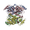
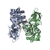
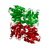
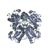
- Assembly
| Deposited unit | 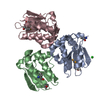
| ||||||||
|---|---|---|---|---|---|---|---|---|---|
| 1 | 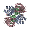
| ||||||||
| 2 |
| ||||||||
| Unit cell |
|
-Components
| #1: Protein | Mass: 13757.277 Da / Num. of mol.: 3 Source method: isolated from a genetically manipulated source Source: (gene. exp.) Planctomyces limnophilus (bacteria) / Strain: DSM 3776 / Gene: Plim_2399 / Plasmid: pMCSG7 / Production host: Escherichia coli (E. coli) / Strain (production host): BL21 (DE3) Magic / References: UniProt: D5SP81#2: Chemical | ChemComp-CL / | Chloride  Mass: 35.453 Da / Num. of mol.: 1 / Source method: obtained synthetically / Formula: Cl Mass: 35.453 Da / Num. of mol.: 1 / Source method: obtained synthetically / Formula: Cl#3: Water | ChemComp-HOH / | Water Mass: 18.015 Da / Num. of mol.: 274 / Source method: isolated from a natural source / Formula: H2O Mass: 18.015 Da / Num. of mol.: 274 / Source method: isolated from a natural source / Formula: H2O |
|---|
-Experimental details
-Experiment
| Experiment | Method: X-RAY DIFFRACTION / Number of used crystals: 1 |
|---|
- Sample preparation
| Crystal | Density Matthews: 2.92 Å3/Da / Density % sol: 57.87 % |
|---|---|
| Crystal grow | Temperature: 289 K / Method: vapor diffusion, sitting drop / pH: 8.5 Details: 0.8M LiCl2, 0.1 Tris:HCl pH8.5, 32% PEG 4K, VAPOR DIFFUSION, SITTING DROP, temperature 289K |
-Data collection
| Diffraction | Mean temperature: 100 K | |||||||||||||||||||||||||||||||||||||||||||||||||||||||||||||||||||||||||||||||||||||||||||||||||||||||||
|---|---|---|---|---|---|---|---|---|---|---|---|---|---|---|---|---|---|---|---|---|---|---|---|---|---|---|---|---|---|---|---|---|---|---|---|---|---|---|---|---|---|---|---|---|---|---|---|---|---|---|---|---|---|---|---|---|---|---|---|---|---|---|---|---|---|---|---|---|---|---|---|---|---|---|---|---|---|---|---|---|---|---|---|---|---|---|---|---|---|---|---|---|---|---|---|---|---|---|---|---|---|---|---|---|---|---|
| Diffraction source | Source: SYNCHROTRON / Site: APS  / Beamline: 19-ID / Wavelength: 0.97935, 0.97945 / Beamline: 19-ID / Wavelength: 0.97935, 0.97945 | |||||||||||||||||||||||||||||||||||||||||||||||||||||||||||||||||||||||||||||||||||||||||||||||||||||||||
| Detector | Type: ADSC QUANTUM 315r / Detector: CCD / Date: Jun 9, 2011 | |||||||||||||||||||||||||||||||||||||||||||||||||||||||||||||||||||||||||||||||||||||||||||||||||||||||||
| Radiation | Monochromator: SAGITALLY FOCUSED Si(111) / Protocol: MAD / Monochromatic (M) / Laue (L): M / Scattering type: x-ray | |||||||||||||||||||||||||||||||||||||||||||||||||||||||||||||||||||||||||||||||||||||||||||||||||||||||||
| Radiation wavelength |
| |||||||||||||||||||||||||||||||||||||||||||||||||||||||||||||||||||||||||||||||||||||||||||||||||||||||||
| Reflection | Resolution: 2.1→50 Å / Num. all: 27534 / Num. obs: 27534 / % possible obs: 97.1 % / Observed criterion σ(I): -3 / Redundancy: 8.7 % / Rmerge(I) obs: 0.101 / Net I/σ(I): 7.2 | |||||||||||||||||||||||||||||||||||||||||||||||||||||||||||||||||||||||||||||||||||||||||||||||||||||||||
| Reflection shell |
|
- Processing
| Software |
| ||||||||||||||||||||||||||||||||||||||||||||||||||||||||||||||||||||||||||||||||||||||||||||||||||||||||||||||||||||||||||||||||||||||||||||||||||||||||||||||||||||||||||
|---|---|---|---|---|---|---|---|---|---|---|---|---|---|---|---|---|---|---|---|---|---|---|---|---|---|---|---|---|---|---|---|---|---|---|---|---|---|---|---|---|---|---|---|---|---|---|---|---|---|---|---|---|---|---|---|---|---|---|---|---|---|---|---|---|---|---|---|---|---|---|---|---|---|---|---|---|---|---|---|---|---|---|---|---|---|---|---|---|---|---|---|---|---|---|---|---|---|---|---|---|---|---|---|---|---|---|---|---|---|---|---|---|---|---|---|---|---|---|---|---|---|---|---|---|---|---|---|---|---|---|---|---|---|---|---|---|---|---|---|---|---|---|---|---|---|---|---|---|---|---|---|---|---|---|---|---|---|---|---|---|---|---|---|---|---|---|---|---|---|---|---|
| Refinement | Method to determine structure: MAD / Resolution: 2.1→50 Å / Cor.coef. Fo:Fc: 0.956 / Cor.coef. Fo:Fc free: 0.925 / Occupancy max: 1 / Occupancy min: 0.5 / SU B: 9.446 / SU ML: 0.114 / SU R Cruickshank DPI: 0.2159 / Cross valid method: THROUGHOUT / ESU R: 0.216 / ESU R Free: 0.186 Stereochemistry target values: MAXIMUM LIKELIHOOD WITH PHASES Details: HYDROGENS HAVE BEEN ADDED IN THE RIDING POSITIONS
| ||||||||||||||||||||||||||||||||||||||||||||||||||||||||||||||||||||||||||||||||||||||||||||||||||||||||||||||||||||||||||||||||||||||||||||||||||||||||||||||||||||||||||
| Solvent computation | Ion probe radii: 0.8 Å / Shrinkage radii: 0.8 Å / VDW probe radii: 1.4 Å / Solvent model: MASK | ||||||||||||||||||||||||||||||||||||||||||||||||||||||||||||||||||||||||||||||||||||||||||||||||||||||||||||||||||||||||||||||||||||||||||||||||||||||||||||||||||||||||||
| Displacement parameters | Biso mean: 27.955 Å2
| ||||||||||||||||||||||||||||||||||||||||||||||||||||||||||||||||||||||||||||||||||||||||||||||||||||||||||||||||||||||||||||||||||||||||||||||||||||||||||||||||||||||||||
| Refinement step | Cycle: LAST / Resolution: 2.1→50 Å
| ||||||||||||||||||||||||||||||||||||||||||||||||||||||||||||||||||||||||||||||||||||||||||||||||||||||||||||||||||||||||||||||||||||||||||||||||||||||||||||||||||||||||||
| Refine LS restraints |
| ||||||||||||||||||||||||||||||||||||||||||||||||||||||||||||||||||||||||||||||||||||||||||||||||||||||||||||||||||||||||||||||||||||||||||||||||||||||||||||||||||||||||||
| LS refinement shell | Resolution: 2.103→2.158 Å / Total num. of bins used: 20
| ||||||||||||||||||||||||||||||||||||||||||||||||||||||||||||||||||||||||||||||||||||||||||||||||||||||||||||||||||||||||||||||||||||||||||||||||||||||||||||||||||||||||||
| Refinement TLS params. | Method: refined / Refine-ID: X-RAY DIFFRACTION
| ||||||||||||||||||||||||||||||||||||||||||||||||||||||||||||||||||||||||||||||||||||||||||||||||||||||||||||||||||||||||||||||||||||||||||||||||||||||||||||||||||||||||||
| Refinement TLS group |
|
