 Movie
Movie Controller

[English] 日本語
 Yorodumi
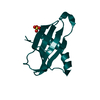
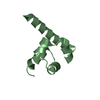
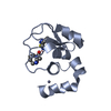
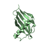
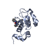
- PDB-3s7r: Crystal structure of a Heterogeneous nuclear ribonucleoprotein A/... -
+ Open data

- Basic information
| Entry | Database: PDB / ID: 3s7r | ||||||
|---|---|---|---|---|---|---|---|
| Title | Crystal structure of a Heterogeneous nuclear ribonucleoprotein A/B (HNRPAB) from HOMO SAPIENS at 2.15 A resolution | ||||||
 Components Components | Heterogeneous nuclear ribonucleoprotein A/B | ||||||
 Keywords Keywords |  RNA BINDING PROTEIN / Ferredoxin-like / Structural Genomics / Joint Center for Structural Genomics / JCSG / Protein Structure Initiative / PSI-BIOLOGY / Partnership for T-Cell Biology / TCELL / Partnership for Stem Cell Biology / STEMCELL RNA BINDING PROTEIN / Ferredoxin-like / Structural Genomics / Joint Center for Structural Genomics / JCSG / Protein Structure Initiative / PSI-BIOLOGY / Partnership for T-Cell Biology / TCELL / Partnership for Stem Cell Biology / STEMCELL | ||||||
| Function / homology |  Function and homology informationepithelial to mesenchymal transition / RNA polymerase II transcription regulator complex / sequence-specific double-stranded DNA binding / sequence-specific DNA binding / ribonucleoprotein complex / mRNA binding / positive regulation of DNA-templated transcription / RNA binding / nucleoplasm / nucleus / cytoplasm Function and homology informationepithelial to mesenchymal transition / RNA polymerase II transcription regulator complex / sequence-specific double-stranded DNA binding / sequence-specific DNA binding / ribonucleoprotein complex / mRNA binding / positive regulation of DNA-templated transcription / RNA binding / nucleoplasm / nucleus / cytoplasmSimilarity search - Function | ||||||
| Biological species |  Homo sapiens (human) Homo sapiens (human) | ||||||
| Method | X-RAY DIFFRACTION / SYNCHROTRON / MAD / Resolution: 2.15 Å | ||||||
 Authors Authors | Joint Center for Structural Genomics (JCSG) / Partnership for T-Cell Biology (TCELL) / Partnership for Stem Cell Biology (STEMCELL) | ||||||
 Citation Citation | Journal: To be published Title: Crystal structure of a Heterogeneous nuclear ribonucleoprotein A/B (HNRPAB) from Homo sapiens at 2.15 A resolution Authors: Joint Center for Structural Genomics (JCSG) / Partnership for T-Cell Biology (TCELL) / Partnership for Stem Cell Biology (STEMCELL) | ||||||
| History |
|
- Structure visualization
| Structure viewer | Molecule: MolmilJmol/JSmol |
|---|
- Downloads & links
-Download
| PDBx/mmCIF format | 3s7r.cif.gz | 76 KB | Display | PDBx/mmCIF format |
|---|---|---|---|---|
| PDB format | pdb3s7r.ent.gz | 59.6 KB | Display | PDB format |
| PDBx/mmJSON format | 3s7r.json.gz | Tree view | PDBx/mmJSON format | |
| Others |  Other downloads Other downloads |
-Validation report
| Arichive directory | https://data.pdbj.org/pub/pdb/validation_reports/s7/3s7rftp://data.pdbj.org/pub/pdb/validation_reports/s7/3s7r | HTTPS FTP |
|---|
-Related structure data
| Similar structure data | |
|---|---|
| Other databases |


-Links
 PDBj
PDBj

- Assembly
| Deposited unit | 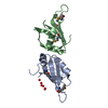
| ||||||||
|---|---|---|---|---|---|---|---|---|---|
| 1 | 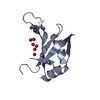
| ||||||||
| 2 | 
| ||||||||
| Unit cell |
|
-Components
| #1: Protein | Mass: 9809.822 Da / Num. of mol.: 2 / Fragment: sequence database residues 59-144 Source method: isolated from a genetically manipulated source Source: (gene. exp.) Homo sapiens (human) / Gene: ABBP1, BC009359, HNRNPAB, HNRPAB / Plasmid: SpeedET / Production host:  Escherichia Coli (E. coli) / Strain (production host): HK100 / References: UniProt: Q99729 Escherichia Coli (E. coli) / Strain (production host): HK100 / References: UniProt: Q99729#2: Chemical | Num. of mol.: 2 / Source method: obtained synthetically #3: Water | ChemComp-HOH / | Water Mass: 18.015 Da / Num. of mol.: 104 / Source method: isolated from a natural source / Formula: H2O Mass: 18.015 Da / Num. of mol.: 104 / Source method: isolated from a natural source / Formula: H2OSequence details | THE CONSTRUCT WAS EXPRESSED WITH AN N-TERMINAL PURIFICATION TAG MGSDKIHHHHHHENLYFQG. THE TAG WAS ...THE CONSTRUCT WAS EXPRESSED WITH AN N-TERMINAL PURIFICATI | |
|---|
-Experimental details
-Experiment
| Experiment | Method: X-RAY DIFFRACTION / Number of used crystals: 1 |
|---|
- Sample preparation
| Crystal | Density Matthews: 2.54 Å3/Da / Density % sol: 51.49 % |
|---|---|
| Crystal grow | Temperature: 277 K / Method: vapor diffusion, sitting drop / pH: 9.5 Details: 40.00% polyethylene glycol 600, 0.1M CHES pH 9.5, NANODROP, VAPOR DIFFUSION, SITTING DROP, temperature 277K |
-Data collection
| Diffraction | Mean temperature: 100 K | |||||||||||||||||||||||||||||||||||||||||||||||||||||||||||||||||||||||||||||
|---|---|---|---|---|---|---|---|---|---|---|---|---|---|---|---|---|---|---|---|---|---|---|---|---|---|---|---|---|---|---|---|---|---|---|---|---|---|---|---|---|---|---|---|---|---|---|---|---|---|---|---|---|---|---|---|---|---|---|---|---|---|---|---|---|---|---|---|---|---|---|---|---|---|---|---|---|---|---|
| Diffraction source | Source: SYNCHROTRON / Site: SSRL  / Beamline: BL9-2 / Wavelength: 0.91162,0.97934,0.97915 / Beamline: BL9-2 / Wavelength: 0.91162,0.97934,0.97915 | |||||||||||||||||||||||||||||||||||||||||||||||||||||||||||||||||||||||||||||
| Detector | Type: MARMOSAIC 325 mm CCD / Detector: CCD / Date: Mar 10, 2011 / Details: double crystal monochromator | |||||||||||||||||||||||||||||||||||||||||||||||||||||||||||||||||||||||||||||
| Radiation | Monochromator: double crystal / Protocol: MAD / Monochromatic (M) / Laue (L): M / Scattering type: x-ray | |||||||||||||||||||||||||||||||||||||||||||||||||||||||||||||||||||||||||||||
| Radiation wavelength |
| |||||||||||||||||||||||||||||||||||||||||||||||||||||||||||||||||||||||||||||
| Reflection | Resolution: 2.15→30.028 Å / Num. obs: 10785 / % possible obs: 97.3 % / Observed criterion σ(I): -3 / Biso Wilson estimate: 31.542 Å2 / Rmerge(I) obs: 0.086 / Net I/σ(I): 7.05 | |||||||||||||||||||||||||||||||||||||||||||||||||||||||||||||||||||||||||||||
| Reflection shell |
|
-Phasing
| Phasing | Method: MAD |
|---|
- Processing
| Software |
| ||||||||||||||||||||||||||||||||||||||||||||||||||||||||||||||||||||||||||||||||||||||||||||||||||||||||||||
|---|---|---|---|---|---|---|---|---|---|---|---|---|---|---|---|---|---|---|---|---|---|---|---|---|---|---|---|---|---|---|---|---|---|---|---|---|---|---|---|---|---|---|---|---|---|---|---|---|---|---|---|---|---|---|---|---|---|---|---|---|---|---|---|---|---|---|---|---|---|---|---|---|---|---|---|---|---|---|---|---|---|---|---|---|---|---|---|---|---|---|---|---|---|---|---|---|---|---|---|---|---|---|---|---|---|---|---|---|---|
| Refinement | Method to determine structure: MAD / Resolution: 2.15→30.028 Å / Cor.coef. Fo:Fc: 0.9508 / Cor.coef. Fo:Fc free: 0.9218 / Occupancy max: 1 / Occupancy min: 0.5 / Cross valid method: THROUGHOUT / σ(F): 0 Details: 1. A MET-INHIBITION PROTOCOL WAS USED FOR SELENOMETHIONINE INCORPORATION DURING PROTEIN EXPRESSION. THE OCCUPANCY OF THE SE ATOMS IN THE MSE RESIDUES WAS REDUCED TO 0.75 FOR THE REDUCED ...Details: 1. A MET-INHIBITION PROTOCOL WAS USED FOR SELENOMETHIONINE INCORPORATION DURING PROTEIN EXPRESSION. THE OCCUPANCY OF THE SE ATOMS IN THE MSE RESIDUES WAS REDUCED TO 0.75 FOR THE REDUCED SCATTERING POWER DUE TO PARTIAL S-MET INCORPORATION. 2. ATOM RECORD CONTAINS SUM OF TLS AND RESIDUAL B FACTORS. ANISOU RECORD CONTAINS SUM OF TLS AND RESIDUAL U FACTORS. 3. NCS RESTRAINTS WERE APPLIED USING BUSTER'S LSSR RESTRAINT REPRESENTATION (-AUTONCS).4.AN UNKNOWN LIGAND (UNL) WAS MODELED AT THE PUTATIVE RNA BINDING SITE.
| ||||||||||||||||||||||||||||||||||||||||||||||||||||||||||||||||||||||||||||||||||||||||||||||||||||||||||||
| Displacement parameters | Biso max: 107.15 Å2 / Biso mean: 40.0225 Å2 / Biso min: 20.31 Å2
| ||||||||||||||||||||||||||||||||||||||||||||||||||||||||||||||||||||||||||||||||||||||||||||||||||||||||||||
| Refinement step | Cycle: LAST / Resolution: 2.15→30.028 Å
| ||||||||||||||||||||||||||||||||||||||||||||||||||||||||||||||||||||||||||||||||||||||||||||||||||||||||||||
| Refine LS restraints |
| ||||||||||||||||||||||||||||||||||||||||||||||||||||||||||||||||||||||||||||||||||||||||||||||||||||||||||||
| LS refinement shell | Resolution: 2.15→2.4 Å / Total num. of bins used: 5
| ||||||||||||||||||||||||||||||||||||||||||||||||||||||||||||||||||||||||||||||||||||||||||||||||||||||||||||
| Refinement TLS params. | Method: refined / Refine-ID: X-RAY DIFFRACTION
| ||||||||||||||||||||||||||||||||||||||||||||||||||||||||||||||||||||||||||||||||||||||||||||||||||||||||||||
| Refinement TLS group | Refine-ID: X-RAY DIFFRACTION / Selection: all
|
