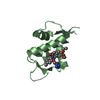
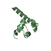
- PDB-6v0m: Sterile alpha-motif from apoptosis signal-regulating kinase 3 -
+
Open data
ID or keywords:
Loading...
-
Basic information
Entry
Database: PDB / ID: 6v0m
Title
Sterile alpha-motif from apoptosis signal-regulating kinase 3
Components
Mitogen-activated protein kinase kinase kinase 15
Keywords
TRANSFERASE / SAM / kinase / ASK
Function / homology
Function and homology information
: / protein serine/threonine kinase activity => GO:0004674 / mitogen-activated protein kinase kinase kinase / MAP kinase kinase kinase activity / protein kinase activity / protein serine kinase activity / ATP binding / metal ion binding Similarity search - Function
MAP3K, TRAFs-binding domain / MAP3K, PH domain / MAP3K TRAFs-binding domain / ASK kinase PH domain / Sterile alpha motif/pointed domain superfamily / Serine/threonine-protein kinase, active site / Serine/Threonine protein kinases active-site signature. / Protein kinase domain / Serine/Threonine protein kinases, catalytic domain / Protein kinase, ATP binding site ...MAP3K, TRAFs-binding domain / MAP3K, PH domain / MAP3K TRAFs-binding domain / ASK kinase PH domain / Sterile alpha motif/pointed domain superfamily / Serine/threonine-protein kinase, active site / Serine/Threonine protein kinases active-site signature. / Protein kinase domain / Serine/Threonine protein kinases, catalytic domain / Protein kinase, ATP binding site / Protein kinases ATP-binding region signature. / Protein kinase domain profile. / Protein kinase domain / Protein kinase-like domain superfamily Similarity search - Domain/homology
Method to determine structure: SAD / Resolution: 1.8→48.13 Å / Cor.coef. Fo:Fc: 0.945 / Cor.coef. Fo:Fc free: 0.92 / SU B: 7.745 / SU ML: 0.116 / Cross valid method: THROUGHOUT / ESU R: 0.144 / ESU R Free: 0.14 Details: U VALUES : WITH TLS ADDED HYDROGENS HAVE BEEN ADDED IN THE RIDING POSITIONS U VALUES : RESIDUAL ONLY
Rfactor
Num. reflection
% reflection
Selection details
Rfree
0.26307
1112
5 %
RANDOM
Rwork
0.21927
-
-
-
obs
0.22145
21264
99.12 %
-
Solvent computation
Ion probe radii: 0.7 Å / Shrinkage radii: 0.7 Å / VDW probe radii: 1.1 Å
Displacement parameters
Biso mean: 37.716 Å2
Baniso -1
Baniso -2
Baniso -3
1-
1.21 Å2
-0 Å2
-0 Å2
2-
-
0.21 Å2
-0 Å2
3-
-
-
-1.42 Å2
Refinement step
Cycle: LAST / Resolution: 1.8→48.13 Å
Protein
Nucleic acid
Ligand
Solvent
Total
Num. atoms
1679
0
0
117
1796
Refine LS restraints
Refine-ID
Type
Dev ideal
Dev ideal target
Number
X-RAY DIFFRACTION
r_bond_refined_d
0.017
0.019
1699
X-RAY DIFFRACTION
r_bond_other_d
0.002
0.02
1652
X-RAY DIFFRACTION
r_angle_refined_deg
1.657
1.928
2284
X-RAY DIFFRACTION
r_angle_other_deg
1.259
2.938
3806
X-RAY DIFFRACTION
r_dihedral_angle_1_deg
5.263
5
204
X-RAY DIFFRACTION
r_dihedral_angle_2_deg
35.567
23.294
85
X-RAY DIFFRACTION
r_dihedral_angle_3_deg
15.216
15
331
X-RAY DIFFRACTION
r_dihedral_angle_4_deg
17.053
15
21
X-RAY DIFFRACTION
r_chiral_restr
0.108
0.2
253
X-RAY DIFFRACTION
r_gen_planes_refined
0.008
0.02
1863
X-RAY DIFFRACTION
r_gen_planes_other
0.001
0.02
358
X-RAY DIFFRACTION
r_mcbond_it
2.637
2.015
825
X-RAY DIFFRACTION
r_mcbond_other
2.629
2.014
824
X-RAY DIFFRACTION
r_mcangle_it
3.363
3.003
1026
X-RAY DIFFRACTION
r_mcangle_other
3.365
3.004
1027
X-RAY DIFFRACTION
r_scbond_it
4.22
2.628
874
X-RAY DIFFRACTION
r_scbond_other
4.213
2.629
872
X-RAY DIFFRACTION
r_scangle_other
6.219
3.723
1258
X-RAY DIFFRACTION
r_long_range_B_refined
7.735
25.995
2102
X-RAY DIFFRACTION
r_long_range_B_other
7.643
25.416
2072
LS refinement shell
Resolution: 1.8→1.843 Å
Rfactor
Num. reflection
% reflection
Rfree
0.376
70
-
Rwork
0.364
1550
-
obs
-
-
98.6 %
Refinement TLS params.
Method: refined / Refine-ID: X-RAY DIFFRACTION
ID
L11 (°2)
L12 (°2)
L13 (°2)
L22 (°2)
L23 (°2)
L33 (°2)
S11 (Å °)
S12 (Å °)
S13 (Å °)
S21 (Å °)
S22 (Å °)
S23 (Å °)
S31 (Å °)
S32 (Å °)
S33 (Å °)
T11 (Å2)
T12 (Å2)
T13 (Å2)
T22 (Å2)
T23 (Å2)
T33 (Å2)
Origin x (Å)
Origin y (Å)
Origin z (Å)
1
4.2788
-0.3121
-0.3014
2.533
-0.2089
1.354
0.0607
-0.0421
0.1472
0.0294
-0.0124
0.0429
-0.0411
0.0798
-0.0483
0.0092
-0.0072
-0.0255
0.0406
-0.0092
0.146
52.971
4.623
66.471
2
5.4105
1.3237
-1.0094
2.8704
0.5716
3.1868
0.1263
-0.3574
0.1417
0.0851
-0.1438
0.1286
-0.161
-0.1693
0.0174
0.0141
0.0053
-0.0063
0.0616
-0.0002
0.1815
29.469
-1.217
77.423
3
3.2129
0.2073
1.2664
2.7249
0.5908
4.8143
-0.1018
-0.1336
0.027
0.0864
-0.0688
0.0029
-0.284
0.147
0.1706
0.0269
0.0021
0.009
0.1002
0.014
0.16
20.749
7.202
97.291
Refinement TLS group
ID
Refine-ID
Refine TLS-ID
Auth asym-ID
Auth seq-ID
1
X-RAY DIFFRACTION
1
A
1238 - 1308
2
X-RAY DIFFRACTION
2
B
1238 - 1308
3
X-RAY DIFFRACTION
3
C
1240 - 1304
+
About Yorodumi
-
News
-
Feb 9, 2022. New format data for meta-information of EMDB entries
New format data for meta-information of EMDB entries
Version 3 of the EMDB header file is now the official format.
The previous official version 1.9 will be removed from the archive.
In the structure databanks used in Yorodumi, some data are registered as the other names, "COVID-19 virus" and "2019-nCoV". Here are the details of the virus and the list of structure data.
Jan 31, 2019. EMDB accession codes are about to change! (news from PDBe EMDB page)
EMDB accession codes are about to change! (news from PDBe EMDB page)
The allocation of 4 digits for EMDB accession codes will soon come to an end. Whilst these codes will remain in use, new EMDB accession codes will include an additional digit and will expand incrementally as the available range of codes is exhausted. The current 4-digit format prefixed with “EMD-” (i.e. EMD-XXXX) will advance to a 5-digit format (i.e. EMD-XXXXX), and so on. It is currently estimated that the 4-digit codes will be depleted around Spring 2019, at which point the 5-digit format will come into force.
The EM Navigator/Yorodumi systems omit the EMD- prefix.
Related info.:Q: What is EMD? / ID/Accession-code notation in Yorodumi/EM Navigator
Yorodumi is a browser for structure data from EMDB, PDB, SASBDB, etc.
This page is also the successor to EM Navigator detail page, and also detail information page/front-end page for Omokage search.
The word "yorodu" (or yorozu) is an old Japanese word meaning "ten thousand". "mi" (miru) is to see.
Related info.:EMDB / PDB / SASBDB / Comparison of 3 databanks / Yorodumi Search / Aug 31, 2016. New EM Navigator & Yorodumi / Yorodumi Papers / Jmol/JSmol / Function and homology information / Changes in new EM Navigator and Yorodumi
 Movie
Movie Controller


 Open data
 Basic information
 Components
 Keywords
 TRANSFERASE / SAM /
  Function and homology information
 Authors
 New Zealand, 1items
  Citation
 Structure visualization
 Downloads & links
 Other downloads


 PDBj
PDBj Assembly
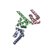


 Mass: 18.015 Da / Num. of mol.: 117 / Source method: isolated from a natural source / Formula: H2O
 Sample preparation
 / Beamline: MX2 / Wavelength: 0.9537, 1.456
 Processing