 Movie
Movie Controller


+ Open data

- Basic information
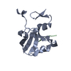
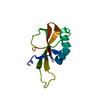
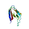
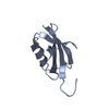
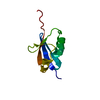
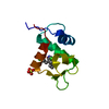
| Entry | Database: PDB / ID: 2qg1 | ||||||
|---|---|---|---|---|---|---|---|
| Title | Crystal structure of the 11th PDZ domain of MPDZ (MUPP1) | ||||||
 Components Components | Multiple PDZ domain protein | ||||||
 Keywords Keywords |  SIGNALING PROTEIN / MPDZ / MUPP1 / Structural Genomics / Structural Genomics Consortium / SGC SIGNALING PROTEIN / MPDZ / MUPP1 / Structural Genomics / Structural Genomics Consortium / SGC | ||||||
| Function / homology |  Function and homology informationtight junction assembly / microtubule organizing center organization / apicolateral plasma membrane / bicellular tight junction / regulation of microtubule cytoskeleton organization / apical part of cell / postsynaptic density / apical plasma membrane / dendrite / plasma membrane / cytoplasm Function and homology informationtight junction assembly / microtubule organizing center organization / apicolateral plasma membrane / bicellular tight junction / regulation of microtubule cytoskeleton organization / apical part of cell / postsynaptic density / apical plasma membrane / dendrite / plasma membrane / cytoplasmSimilarity search - Function | ||||||
| Biological species |  Homo sapiens (human) Homo sapiens (human) | ||||||
| Method | X-RAY DIFFRACTION / SYNCHROTRON / MOLECULAR REPLACEMENT / Resolution: 1.4 Å | ||||||
 Authors Authors | Papagrigoriou, E. / Salah, E. / Phillips, C. / Savitsky, P. / Boisguerin, P. / Oschkinat, H. / Gileadi, C. / Yang, X. / Elkins, J.M. / Ugochukwu, E. ...Papagrigoriou, E. / Salah, E. / Phillips, C. / Savitsky, P. / Boisguerin, P. / Oschkinat, H. / Gileadi, C. / Yang, X. / Elkins, J.M. / Ugochukwu, E. / Bunkoczi, G. / Uppenberg, J. / Sundstrom, M. / Arrowsmith, C.H. / Weigelt, J. / Edwards, A. / von Delft, F. / Doyle, D. / Structural Genomics Consortium (SGC) | ||||||
 Citation Citation | Journal: To be Published Title: Crystal structure of the 11th PDZ domain of MPDZ (MUPP1). Authors: Papagrigoriou, E. / Salah, E. / Phillips, C. / Savitsky, P. / Boisguerin, P. / Oschkinat, H. / Gileadi, C. / Yang, X. / Elkins, J.M. / Ugochukwu, E. / Bunkoczi, G. / Uppenberg, J. / Doyle, D. | ||||||
| History |
|
- Structure visualization
| Structure viewer | Molecule: MolmilJmol/JSmol |
|---|
- Downloads & links
-Download
| PDBx/mmCIF format | 2qg1.cif.gz | 52.8 KB | Display | PDBx/mmCIF format |
|---|---|---|---|---|
| PDB format | pdb2qg1.ent.gz | 37.9 KB | Display | PDB format |
| PDBx/mmJSON format | 2qg1.json.gz | Tree view | PDBx/mmJSON format | |
| Others |  Other downloads Other downloads |
-Validation report
| Arichive directory | https://data.pdbj.org/pub/pdb/validation_reports/qg/2qg1ftp://data.pdbj.org/pub/pdb/validation_reports/qg/2qg1 | HTTPS FTP |
|---|
-Related structure data


-Links
 PDBj
PDBj- Assembly
| Deposited unit | 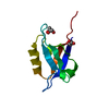
| ||||||||
|---|---|---|---|---|---|---|---|---|---|
| 1 |
| ||||||||
| Unit cell |
|
-Components
| #1: Protein | Mass: 9574.009 Da / Num. of mol.: 1 / Fragment: 11th PDZ domain / Mutation: E1727G Source method: isolated from a genetically manipulated source Source: (gene. exp.) Homo sapiens (human) / Gene: MPDZ, MUPP1 / Plasmid: pNIC28-Bsa4 / Production host:  Escherichia coli (E. coli) / Strain (production host): BL21(DE3)-R3-pRARE2 / References: UniProt: O75970 Escherichia coli (E. coli) / Strain (production host): BL21(DE3)-R3-pRARE2 / References: UniProt: O75970 | ||
|---|---|---|---|
| #2: Chemical | Ethylene glycol  Mass: 62.068 Da / Num. of mol.: 2 / Source method: obtained synthetically / Formula: C2H6O2 Mass: 62.068 Da / Num. of mol.: 2 / Source method: obtained synthetically / Formula: C2H6O2#3: Water | ChemComp-HOH / | Water Mass: 18.015 Da / Num. of mol.: 120 / Source method: isolated from a natural source / Formula: H2O Mass: 18.015 Da / Num. of mol.: 120 / Source method: isolated from a natural source / Formula: H2O |
-Experimental details
-Experiment
| Experiment | Method: X-RAY DIFFRACTION / Number of used crystals: 1 |
|---|
- Sample preparation
| Crystal | Density Matthews: 2.05 Å3/Da / Density % sol: 39.96 % |
|---|---|
| Crystal grow | Temperature: 293 K / Method: vapor diffusion, sitting drop / pH: 4 Details: 0.8M (NH4)2SO4, 0.1M Citrate pH 4.0, VAPOR DIFFUSION, SITTING DROP, temperature 293K |
-Data collection
| Diffraction | Mean temperature: 100 K |
|---|---|
| Diffraction source | Source: SYNCHROTRON / Site: SLS  / Beamline: X10SA / Wavelength: 0.9182 Å / Beamline: X10SA / Wavelength: 0.9182 Å |
| Detector | Type: MARMOSAIC 225 mm CCD / Detector: CCD / Date: Feb 11, 2007 |
| Radiation | Protocol: SINGLE WAVELENGTH / Monochromatic (M) / Laue (L): M / Scattering type: x-ray |
| Radiation wavelength | Wavelength: 0.9182 Å / Relative weight: 1 |
| Reflection | Resolution: 1.4→31.8 Å / Num. all: 15501 / Num. obs: 15501 / % possible obs: 96.6 % / Observed criterion σ(F): 0 / Observed criterion σ(I): 0 / Redundancy: 4.6 % / Rmerge(I) obs: 0.073 / Rsym value: 0.073 / Net I/σ(I): 13.5 |
| Reflection shell | Resolution: 1.4→1.48 Å / Redundancy: 3.8 % / Rmerge(I) obs: 0.394 / Mean I/σ(I) obs: 2.6 / Num. unique all: 8319 / Rsym value: 0.394 / % possible all: 94.7 |
- Processing
| Software |
| ||||||||||||||||||||||||||||||||||||||||||||||||||||||||||||||||||||||||||||||||||||||||||||||||||||||||||||||||||||||||||||||||||||||||||||
|---|---|---|---|---|---|---|---|---|---|---|---|---|---|---|---|---|---|---|---|---|---|---|---|---|---|---|---|---|---|---|---|---|---|---|---|---|---|---|---|---|---|---|---|---|---|---|---|---|---|---|---|---|---|---|---|---|---|---|---|---|---|---|---|---|---|---|---|---|---|---|---|---|---|---|---|---|---|---|---|---|---|---|---|---|---|---|---|---|---|---|---|---|---|---|---|---|---|---|---|---|---|---|---|---|---|---|---|---|---|---|---|---|---|---|---|---|---|---|---|---|---|---|---|---|---|---|---|---|---|---|---|---|---|---|---|---|---|---|---|---|---|
| Refinement | Method to determine structure: MOLECULAR REPLACEMENT Starting model: PDB entries 2HE2, 1TP3 Resolution: 1.4→31.8 Å / Cor.coef. Fo:Fc: 0.969 / Cor.coef. Fo:Fc free: 0.952 / SU B: 3.633 / SU ML: 0.064 / Cross valid method: THROUGHOUT / σ(F): 0 / ESU R: 0.08 / ESU R Free: 0.079 / Stereochemistry target values: MAXIMUM LIKELIHOOD / Details: HYDROGENS HAVE BEEN ADDED IN THE RIDING POSITIONS
| ||||||||||||||||||||||||||||||||||||||||||||||||||||||||||||||||||||||||||||||||||||||||||||||||||||||||||||||||||||||||||||||||||||||||||||
| Solvent computation | Ion probe radii: 0.8 Å / Shrinkage radii: 0.8 Å / VDW probe radii: 1.2 Å / Solvent model: BABINET MODEL WITH MASK | ||||||||||||||||||||||||||||||||||||||||||||||||||||||||||||||||||||||||||||||||||||||||||||||||||||||||||||||||||||||||||||||||||||||||||||
| Displacement parameters | Biso mean: 14.93 Å2
| ||||||||||||||||||||||||||||||||||||||||||||||||||||||||||||||||||||||||||||||||||||||||||||||||||||||||||||||||||||||||||||||||||||||||||||
| Refinement step | Cycle: LAST / Resolution: 1.4→31.8 Å
| ||||||||||||||||||||||||||||||||||||||||||||||||||||||||||||||||||||||||||||||||||||||||||||||||||||||||||||||||||||||||||||||||||||||||||||
| Refine LS restraints |
| ||||||||||||||||||||||||||||||||||||||||||||||||||||||||||||||||||||||||||||||||||||||||||||||||||||||||||||||||||||||||||||||||||||||||||||
| LS refinement shell | Resolution: 1.4→1.436 Å / Total num. of bins used: 20
|
