 Movie
Movie Controller

[English] 日本語
 Yorodumi
- PDB-3rp7: Crystal Structure of Klebsiella pneumoniae HpxO complexed with FA... -
+ Open data

- Basic information
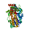
| Entry | Database: PDB / ID: 3rp7 | ||||||
|---|---|---|---|---|---|---|---|
| Title | Crystal Structure of Klebsiella pneumoniae HpxO complexed with FAD and uric acid | ||||||
 Components Components | flavoprotein monooxygenase | ||||||
 Keywords Keywords |  OXIDOREDUCTASE / FAD-binding protein / monooxygenase OXIDOREDUCTASE / FAD-binding protein / monooxygenase | ||||||
| Function / homology |  Function and homology informationFAD-dependent urate hydroxylase / FAD-dependent urate hydroxylase activity / oxidoreductase activity, acting on paired donors, with incorporation or reduction of molecular oxygen, NAD(P)H as one donor, and incorporation of one atom of oxygen / urate oxidase activity / urate catabolic process / purine nucleobase metabolic process / FAD binding Function and homology informationFAD-dependent urate hydroxylase / FAD-dependent urate hydroxylase activity / oxidoreductase activity, acting on paired donors, with incorporation or reduction of molecular oxygen, NAD(P)H as one donor, and incorporation of one atom of oxygen / urate oxidase activity / urate catabolic process / purine nucleobase metabolic process / FAD bindingSimilarity search - Function | ||||||
| Biological species |  Klebsiella pneumoniae (bacteria) Klebsiella pneumoniae (bacteria) | ||||||
| Method | X-RAY DIFFRACTION / SYNCHROTRON / FOURIER SYNTHESIS / Resolution: 2.042 Å | ||||||
 Authors Authors | Hicks, K.A. / O'Leary, S.E. / Begley, T.P. / Ealick, S.E. | ||||||
 Citation Citation | Journal: Biochemistry / Year: 2013 Title: Structural and Mechanistic Studies of HpxO, a Novel Flavin Adenine Dinucleotide-Dependent Urate Oxidase from Klebsiella pneumoniae. Authors: Hicks, K.A. / O'Leary, S.E. / Begley, T.P. / Ealick, S.E. | ||||||
| History |
|
- Structure visualization
| Structure viewer | Molecule: MolmilJmol/JSmol |
|---|
- Downloads & links
-Download
| PDBx/mmCIF format | 3rp7.cif.gz | 90.4 KB | Display | PDBx/mmCIF format |
|---|---|---|---|---|
| PDB format | pdb3rp7.ent.gz | 70.6 KB | Display | PDB format |
| PDBx/mmJSON format | 3rp7.json.gz | Tree view | PDBx/mmJSON format | |
| Others |  Other downloads Other downloads |
-Validation report
| Arichive directory | https://data.pdbj.org/pub/pdb/validation_reports/rp/3rp7ftp://data.pdbj.org/pub/pdb/validation_reports/rp/3rp7 | HTTPS FTP |
|---|
-Related structure data


-Links
 PDBj
PDBj- Assembly
| Deposited unit | 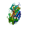
| ||||||||
|---|---|---|---|---|---|---|---|---|---|
| 1 |
| ||||||||
| Unit cell |
|
-Components
| #1: Protein | Mass: 45401.504 Da / Num. of mol.: 1 Source method: isolated from a genetically manipulated source Source: (gene. exp.) Klebsiella pneumoniae (bacteria) / Strain: ATCC 700721 / MGH 78578 / Gene: KPN78578_16330, KPN_01663 / Plasmid: modified pET28a / Production host: Escherichia coli (E. coli) / Strain (production host): B834(DE3) / References: UniProt: A6T923 |
|---|---|
| #2: Chemical | ChemComp-URC / Uric acid  Mass: 168.110 Da / Num. of mol.: 1 / Source method: obtained synthetically / Formula: C5H4N4O3 Mass: 168.110 Da / Num. of mol.: 1 / Source method: obtained synthetically / Formula: C5H4N4O3 |
| #3: Chemical | ChemComp-FAD / Flavin adenine dinucleotide  Mass: 785.550 Da / Num. of mol.: 1 / Source method: obtained synthetically / Formula: C27H33N9O15P2 / Comment: FAD*YM Mass: 785.550 Da / Num. of mol.: 1 / Source method: obtained synthetically / Formula: C27H33N9O15P2 / Comment: FAD*YM |
| #4: Water | ChemComp-HOH / Water Mass: 18.015 Da / Num. of mol.: 194 / Source method: isolated from a natural source / Formula: H2O Mass: 18.015 Da / Num. of mol.: 194 / Source method: isolated from a natural source / Formula: H2O |
-Experimental details
-Experiment
| Experiment | Method: X-RAY DIFFRACTION / Number of used crystals: 1 |
|---|
- Sample preparation
| Crystal | Density Matthews: 2.06 Å3/Da / Density % sol: 40.26 % |
|---|---|
| Crystal grow | Temperature: 291 K / Method: hanging drop Details: 0.05 M KH2PO4, 20% PEG 8000, hanging drop, temperature 291K |
-Data collection
| Diffraction | Mean temperature: 100 K | |||||||||||||||||||||||||||||||||||||||||||||||||||||||||||||||||||||||||||||
|---|---|---|---|---|---|---|---|---|---|---|---|---|---|---|---|---|---|---|---|---|---|---|---|---|---|---|---|---|---|---|---|---|---|---|---|---|---|---|---|---|---|---|---|---|---|---|---|---|---|---|---|---|---|---|---|---|---|---|---|---|---|---|---|---|---|---|---|---|---|---|---|---|---|---|---|---|---|---|
| Diffraction source | Source: SYNCHROTRON / Site: APS  / Beamline: 24-ID-C / Wavelength: 0.9791 Å / Beamline: 24-ID-C / Wavelength: 0.9791 Å | |||||||||||||||||||||||||||||||||||||||||||||||||||||||||||||||||||||||||||||
| Detector | Type: ADSC QUANTUM 315 / Detector: CCD / Date: Aug 18, 2009 | |||||||||||||||||||||||||||||||||||||||||||||||||||||||||||||||||||||||||||||
| Radiation | Monochromator: Cryo-Cooled Si(111) double crystal / Protocol: SINGLE WAVELENGTH / Monochromatic (M) / Laue (L): M / Scattering type: x-ray | |||||||||||||||||||||||||||||||||||||||||||||||||||||||||||||||||||||||||||||
| Radiation wavelength | Wavelength: 0.9791 Å / Relative weight: 1 | |||||||||||||||||||||||||||||||||||||||||||||||||||||||||||||||||||||||||||||
| Reflection | Resolution: 2.04→50 Å / Num. all: 24401 / Num. obs: 24401 / % possible obs: 99.7 % / Observed criterion σ(I): -3 / Redundancy: 5.5 % / Rmerge(I) obs: 0.093 / Χ2: 5.912 / Net I/σ(I): 22.8 | |||||||||||||||||||||||||||||||||||||||||||||||||||||||||||||||||||||||||||||
| Reflection shell |
|
- Processing
| Software |
| ||||||||||||||||||||||||||||||||||||||||||||||||||||||||||||||||||||||
|---|---|---|---|---|---|---|---|---|---|---|---|---|---|---|---|---|---|---|---|---|---|---|---|---|---|---|---|---|---|---|---|---|---|---|---|---|---|---|---|---|---|---|---|---|---|---|---|---|---|---|---|---|---|---|---|---|---|---|---|---|---|---|---|---|---|---|---|---|---|---|---|
| Refinement | Method to determine structure: FOURIER SYNTHESIS / Resolution: 2.042→33.92 Å / Occupancy max: 1 / Occupancy min: 1 / FOM work R set: 0.8481 / SU ML: 0.21 / σ(F): 1.34 / Phase error: 21.83 / Stereochemistry target values: ML
| ||||||||||||||||||||||||||||||||||||||||||||||||||||||||||||||||||||||
| Solvent computation | Shrinkage radii: 0.95 Å / VDW probe radii: 1.2 Å / Solvent model: FLAT BULK SOLVENT MODEL / Bsol: 39.888 Å2 / ksol: 0.364 e/Å3 | ||||||||||||||||||||||||||||||||||||||||||||||||||||||||||||||||||||||
| Displacement parameters | Biso max: 63.31 Å2 / Biso mean: 22.8971 Å2 / Biso min: 9.52 Å2
| ||||||||||||||||||||||||||||||||||||||||||||||||||||||||||||||||||||||
| Refinement step | Cycle: LAST / Resolution: 2.042→33.92 Å
| ||||||||||||||||||||||||||||||||||||||||||||||||||||||||||||||||||||||
| Refine LS restraints |
| ||||||||||||||||||||||||||||||||||||||||||||||||||||||||||||||||||||||
| LS refinement shell | Refine-ID: X-RAY DIFFRACTION / Total num. of bins used: 9
|
