 Movie
Movie Controller

[English] 日本語
 Yorodumi
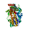
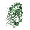
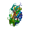
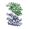
- PDB-5ikd: Asymmetric sulfoxidation by engineering the heme pocket of a dye-... -
+ Open data

- Basic information
| Entry | Database: PDB / ID: 5ikd | ||||||||||||
|---|---|---|---|---|---|---|---|---|---|---|---|---|---|
| Title | Asymmetric sulfoxidation by engineering the heme pocket of a dye-decolorizing peroxidase | ||||||||||||
 Components Components | Dye-decolorizing peroxidase | ||||||||||||
 Keywords Keywords |  OXIDOREDUCTASE / DECOLORIZING PEROXIDASE (DYP) / F359G VARIANT / HEME OXIDOREDUCTASE / DECOLORIZING PEROXIDASE (DYP) / F359G VARIANT / HEME | ||||||||||||
| Function / homology |  Function and homology informationdye decolorizing peroxidase / lactoperoxidase activity / peroxidase / peroxidase activity / heme binding / extracellular region / metal ion binding Function and homology informationdye decolorizing peroxidase / lactoperoxidase activity / peroxidase / peroxidase activity / heme binding / extracellular region / metal ion bindingSimilarity search - Function | ||||||||||||
| Biological species |  Auricularia auricula-judae (ear fungus) Auricularia auricula-judae (ear fungus) | ||||||||||||
| Method | X-RAY DIFFRACTION / SYNCHROTRON / MOLECULAR REPLACEMENT / Resolution: 1.109 Å | ||||||||||||
 Authors Authors | Romero, A. / Davo-Siguero, I. / Martinez, A.T. | ||||||||||||
| Funding support |  Spain, 1items Spain, 1items
| ||||||||||||
 Citation Citation | Journal: Catalysis Science And Technology / Year: 2016 Title: Asymmetric sulfoxidation by engineering the heme pocket of a dye-decolorizing peroxidase Authors: Linde, D. / Canellas, M. / Davo-Siguero, I. / Romero, A. / Lucas, F. / Ruiz-Duenas, F.J. / Guallar, V. / Martinez, A.T. #1: Journal: Biochem. J. / Year: 2015Title: Catalytic surface radical in dye-decolorizing peroxidase: a computational, spectroscopic and site-directed mutagenesis study. Authors: Pogni, R. / Canellas, M. / Lucas, F. / Guallar, V. / Baratto, M.C. / Sinicropi, A. / Saez-Jimenez, V. / Coscolin, C. / Romero, A. / Medrano, F.J. / Ruiz-Duenas, F.J. / Martinez, A.T. | ||||||||||||
| History |
|
- Structure visualization
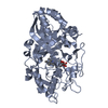
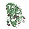
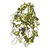
| Structure viewer | Molecule: MolmilJmol/JSmol |
|---|
- Downloads & links
-Download
| PDBx/mmCIF format | 5ikd.cif.gz | 260.6 KB | Display | PDBx/mmCIF format |
|---|---|---|---|---|
| PDB format | pdb5ikd.ent.gz | 210.4 KB | Display | PDB format |
| PDBx/mmJSON format | 5ikd.json.gz | Tree view | PDBx/mmJSON format | |
| Others |  Other downloads Other downloads |
-Validation report
| Arichive directory | https://data.pdbj.org/pub/pdb/validation_reports/ik/5ikdftp://data.pdbj.org/pub/pdb/validation_reports/ik/5ikd | HTTPS FTP |
|---|
-Related structure data
| Related structure data | 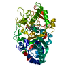 5ikgC  4w7jS S: Starting model for refinement C: citing same article ( |
|---|---|
| Similar structure data |


-Links
 PDBj
PDBj- Assembly
| Deposited unit | 
| |||||||||
|---|---|---|---|---|---|---|---|---|---|---|
| 1 |
| |||||||||
| Unit cell |
| |||||||||
| Components on special symmetry positions |
|
-Components
| #1: Protein | Mass: 46713.805 Da / Num. of mol.: 1 / Fragment: UNP residues 64-509 / Mutation: F359G Source method: isolated from a genetically manipulated source Source: (gene. exp.) Auricularia auricula-judae (ear fungus)Gene: dyp1 / Production host:  Escherichia coli (E. coli) / References: UniProt: I2DBY1, dye decolorizing peroxidase Escherichia coli (E. coli) / References: UniProt: I2DBY1, dye decolorizing peroxidase |
|---|---|
| #2: Chemical | ChemComp-HEM / Heme B  Mass: 616.487 Da / Num. of mol.: 1 / Source method: obtained synthetically / Formula: C34H32FeN4O4 Mass: 616.487 Da / Num. of mol.: 1 / Source method: obtained synthetically / Formula: C34H32FeN4O4 |
| #3: Water | ChemComp-HOH / Water Mass: 18.015 Da / Num. of mol.: 576 / Source method: isolated from a natural source / Formula: H2O Mass: 18.015 Da / Num. of mol.: 576 / Source method: isolated from a natural source / Formula: H2O |
-Experimental details
-Experiment
| Experiment | Method: X-RAY DIFFRACTION / Number of used crystals: 1 |
|---|
- Sample preparation
| Crystal | Density Matthews: 2.58 Å3/Da / Density % sol: 52.41 % |
|---|---|
| Crystal grow | Temperature: 295 K / Method: vapor diffusion, sitting drop / Details: 2M magnesium formate and 20% PEG 3350 |
-Data collection
| Diffraction | Mean temperature: 100 K | |||||||||||||||
|---|---|---|---|---|---|---|---|---|---|---|---|---|---|---|---|---|
| Diffraction source | Source: SYNCHROTRON / Site: ALBA / Beamline: XALOC / Wavelength: 0.9795 Å | |||||||||||||||
| Detector | Type: DECTRIS PILATUS3 6M / Detector: PIXEL / Date: Mar 24, 2015 | |||||||||||||||
| Radiation | Protocol: SINGLE WAVELENGTH / Monochromatic (M) / Laue (L): M / Scattering type: x-ray | |||||||||||||||
| Radiation wavelength | Wavelength: 0.9795 Å / Relative weight: 1 | |||||||||||||||
| Reflection | Resolution: 1.109→46.63 Å / Num. obs: 184770 / % possible obs: 98.4 % / Redundancy: 5.4 % / Biso Wilson estimate: 9.14 Å2 / CC1/2: 0.997 / Rmerge(I) obs: 0.097 / Net I/σ(I): 9.5 | |||||||||||||||
| Reflection shell |
|
- Processing
| Software |
| |||||||||||||||||||||||||||||||||||||||||||||||||||||||||||||||||||||||||||||||||||||||||||||||||||||||||
|---|---|---|---|---|---|---|---|---|---|---|---|---|---|---|---|---|---|---|---|---|---|---|---|---|---|---|---|---|---|---|---|---|---|---|---|---|---|---|---|---|---|---|---|---|---|---|---|---|---|---|---|---|---|---|---|---|---|---|---|---|---|---|---|---|---|---|---|---|---|---|---|---|---|---|---|---|---|---|---|---|---|---|---|---|---|---|---|---|---|---|---|---|---|---|---|---|---|---|---|---|---|---|---|---|---|---|
| Refinement | Method to determine structure: MOLECULAR REPLACEMENT Starting model: 4W7J Resolution: 1.109→41.261 Å / SU ML: 0.1 / Cross valid method: FREE R-VALUE / σ(F): 1.34 / Phase error: 15.48
| |||||||||||||||||||||||||||||||||||||||||||||||||||||||||||||||||||||||||||||||||||||||||||||||||||||||||
| Solvent computation | Shrinkage radii: 0.9 Å / VDW probe radii: 1.11 Å | |||||||||||||||||||||||||||||||||||||||||||||||||||||||||||||||||||||||||||||||||||||||||||||||||||||||||
| Displacement parameters | Biso max: 79.87 Å2 / Biso mean: 14.0754 Å2 / Biso min: 5.01 Å2 | |||||||||||||||||||||||||||||||||||||||||||||||||||||||||||||||||||||||||||||||||||||||||||||||||||||||||
| Refinement step | Cycle: final / Resolution: 1.109→41.261 Å
| |||||||||||||||||||||||||||||||||||||||||||||||||||||||||||||||||||||||||||||||||||||||||||||||||||||||||
| Refine LS restraints |
| |||||||||||||||||||||||||||||||||||||||||||||||||||||||||||||||||||||||||||||||||||||||||||||||||||||||||
| LS refinement shell | Refine-ID: X-RAY DIFFRACTION / Total num. of bins used: 14
|
