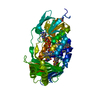
 Movie
Movie Controller

[English] 日本語
 Yorodumi
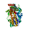
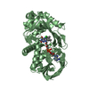
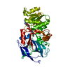
- PDB-3hhc: Interferon-lambda is functionally an interferon but structurally ... -
+ Open data

- Basic information
| Entry | Database: PDB / ID: 3hhc | ||||||
|---|---|---|---|---|---|---|---|
| Title | Interferon-lambda is functionally an interferon but structurally related to the IL-10 family | ||||||
 Components Components | Interleukin-28B | ||||||
 Keywords Keywords |  CYTOKINE / Interferon / IL-22 / antiviral / Antiviral defense / Secreted CYTOKINE / Interferon / IL-22 / antiviral / Antiviral defense / Secreted | ||||||
| Function / homology |  Function and homology information Function and homology informationtype III interferon-mediated signaling pathway / negative regulation of viral genome replication / Interleukin-20 family signaling / cell surface receptor signaling pathway via JAK-STAT / cytokine activity / cellular response to virus / positive regulation of immune response / defense response to virus / signaling receptor binding / innate immune response ...type III interferon-mediated signaling pathway / negative regulation of viral genome replication / Interleukin-20 family signaling / cell surface receptor signaling pathway via JAK-STAT / cytokine activity / cellular response to virus / positive regulation of immune response / defense response to virus / signaling receptor binding / innate immune response / extracellular space / extracellular regionSimilarity search - Function | ||||||
| Biological species |  Homo sapiens (human) Homo sapiens (human) | ||||||
| Method | X-RAY DIFFRACTION / SYNCHROTRON / MIR / Resolution: 2.8 Å | ||||||
 Authors Authors | Gad, H.H. / Hamming, O.J. / Hartmann, R. | ||||||
 Citation Citation | Journal: J.Biol.Chem. / Year: 2009 Title: Interferon-{lambda} Is Functionally an Interferon but Structurally Related to the Interleukin-10 Family Authors: Gad, H.H. / Dellgren, C. / Hamming, O.J. / Vends, S. / Paludan, S.R. / Hartmann, R. | ||||||
| History |
|
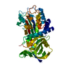
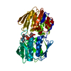
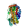
- Structure visualization
| Structure viewer | Molecule: MolmilJmol/JSmol |
|---|
- Downloads & links
-Download
| PDBx/mmCIF format | 3hhc.cif.gz | 239.3 KB | Display | PDBx/mmCIF format |
|---|---|---|---|---|
| PDB format | pdb3hhc.ent.gz | 202.1 KB | Display | PDB format |
| PDBx/mmJSON format | 3hhc.json.gz | Tree view | PDBx/mmJSON format | |
| Others |  Other downloads Other downloads |
-Validation report
| Arichive directory | https://data.pdbj.org/pub/pdb/validation_reports/hh/3hhcftp://data.pdbj.org/pub/pdb/validation_reports/hh/3hhc | HTTPS FTP |
|---|
-Related structure data
| Similar structure data |
|---|


-Links
 PDBj
PDBj

- Assembly
| Deposited unit | 
| ||||||||
|---|---|---|---|---|---|---|---|---|---|
| 1 | 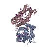
| ||||||||
| 2 | 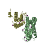
| ||||||||
| Unit cell |
|
-Components
| #1: Protein | Mass: 21738.408 Da / Num. of mol.: 4 Source method: isolated from a genetically manipulated source Source: (gene. exp.) Homo sapiens (human) / Gene: IL28B, IFNL3, IFNL4, IL28C, ZCYTO22 / Production host:  Escherichia coli (E. coli) / References: UniProt: Q8IZI9 Escherichia coli (E. coli) / References: UniProt: Q8IZI9#2: Chemical | ChemComp-IOD / Iodide  Mass: 126.904 Da / Num. of mol.: 35 / Source method: obtained synthetically / Formula: I Mass: 126.904 Da / Num. of mol.: 35 / Source method: obtained synthetically / Formula: I#3: Water | ChemComp-HOH / | Water Mass: 18.015 Da / Num. of mol.: 57 / Source method: isolated from a natural source / Formula: H2O Mass: 18.015 Da / Num. of mol.: 57 / Source method: isolated from a natural source / Formula: H2O |
|---|
-Experimental details
-Experiment
| Experiment | Method: X-RAY DIFFRACTION / Number of used crystals: 1 |
|---|
- Sample preparation
| Crystal | Density Matthews: 2.07 Å3/Da / Density % sol: 40.58 % |
|---|---|
| Crystal grow | Temperature: 285 K / Method: vapor diffusion, sitting drop / pH: 5.5 Details: 28 % w/v PEG 4000, 200 mM ammonium sulfate, 3 % v/v isopropyl alcohol , pH 5.5, VAPOR DIFFUSION, SITTING DROP, temperature 285K |
-Data collection
| Diffraction |
| ||||||||||||||||||||||||||||||||||||||||||||||||||||||||||||||||||
|---|---|---|---|---|---|---|---|---|---|---|---|---|---|---|---|---|---|---|---|---|---|---|---|---|---|---|---|---|---|---|---|---|---|---|---|---|---|---|---|---|---|---|---|---|---|---|---|---|---|---|---|---|---|---|---|---|---|---|---|---|---|---|---|---|---|---|---|
| Diffraction source |
| ||||||||||||||||||||||||||||||||||||||||||||||||||||||||||||||||||
| Detector |
| ||||||||||||||||||||||||||||||||||||||||||||||||||||||||||||||||||
| Radiation | Protocol: SINGLE WAVELENGTH / Monochromatic (M) / Laue (L): M / Scattering type: x-ray | ||||||||||||||||||||||||||||||||||||||||||||||||||||||||||||||||||
| Radiation wavelength |
| ||||||||||||||||||||||||||||||||||||||||||||||||||||||||||||||||||
| Reflection | Resolution: 2.8→25 Å / Num. obs: 39210 / % possible obs: 91.3 % / Redundancy: 5.8 % / Rmerge(I) obs: 0.062 / Χ2: 1.062 / Net I/σ(I): 24.389 | ||||||||||||||||||||||||||||||||||||||||||||||||||||||||||||||||||
| Reflection shell |
|


- Processing
| Software |
| ||||||||||||||||||||||||||||||||||||||||||||||||||||||||||||||||||||||
|---|---|---|---|---|---|---|---|---|---|---|---|---|---|---|---|---|---|---|---|---|---|---|---|---|---|---|---|---|---|---|---|---|---|---|---|---|---|---|---|---|---|---|---|---|---|---|---|---|---|---|---|---|---|---|---|---|---|---|---|---|---|---|---|---|---|---|---|---|---|---|---|
| Refinement | Method to determine structure: MIR / Resolution: 2.8→23.863 Å / Occupancy max: 1 / Occupancy min: 0 / FOM work R set: 0.718 / SU ML: 1.23 / σ(F): 1.33 / Stereochemistry target values: ML
| ||||||||||||||||||||||||||||||||||||||||||||||||||||||||||||||||||||||
| Solvent computation | Shrinkage radii: 0.9 Å / VDW probe radii: 1.11 Å / Solvent model: FLAT BULK SOLVENT MODEL / Bsol: 54.646 Å2 / ksol: 0.268 e/Å3 | ||||||||||||||||||||||||||||||||||||||||||||||||||||||||||||||||||||||
| Displacement parameters | Biso max: 330.34 Å2 / Biso mean: 94.783 Å2 / Biso min: 24.66 Å2
| ||||||||||||||||||||||||||||||||||||||||||||||||||||||||||||||||||||||
| Refinement step | Cycle: LAST / Resolution: 2.8→23.863 Å
| ||||||||||||||||||||||||||||||||||||||||||||||||||||||||||||||||||||||
| Refine LS restraints |
| ||||||||||||||||||||||||||||||||||||||||||||||||||||||||||||||||||||||
| LS refinement shell | Refine-ID: X-RAY DIFFRACTION / Total num. of bins used: 9
| ||||||||||||||||||||||||||||||||||||||||||||||||||||||||||||||||||||||
| Refinement TLS params. | Method: refined / Origin x: -15.0892 Å / Origin y: 27.2206 Å / Origin z: 64.5259 Å
| ||||||||||||||||||||||||||||||||||||||||||||||||||||||||||||||||||||||
| Refinement TLS group |
|
