- PDB-3qt2: Structure of a cytokine ligand-receptor complex -
+
Open data
ID or keywords:
Loading...
-
Basic information
Entry
Database: PDB / ID: 3qt2
Title
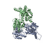
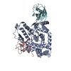
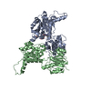
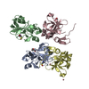
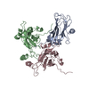
Structure of a cytokine ligand-receptor complex
Components
Interleukin-5 receptor subunit alpha
Interleukin-5Interleukin 5
Keywords
PROTEIN BINDING/IMMUNE SYSTEM / cytokine type I receptor fold / fibronectin type III modules / four-helical bundle / Cytokine / Ligand-receptor complex / membrane / PROTEIN BINDING-IMMUNE SYSTEM complex
Function / homology
Function and homology information
interleukin-5 receptor activity / regulation of interleukin-5 production / positive regulation of eosinophil differentiation / interleukin-5 receptor binding / interleukin-5-mediated signaling pathway / positive regulation of leukocyte proliferation / inflammatory response to antigenic stimulus / positive regulation of podosome assembly / cytokine receptor activity / positive regulation of immunoglobulin production ...interleukin-5 receptor activity / regulation of interleukin-5 production / positive regulation of eosinophil differentiation / interleukin-5 receptor binding / interleukin-5-mediated signaling pathway / positive regulation of leukocyte proliferation / inflammatory response to antigenic stimulus / positive regulation of podosome assembly / cytokine receptor activity / positive regulation of immunoglobulin production / cytokine binding / Interleukin-3, Interleukin-5 and GM-CSF signaling / Interleukin receptor SHC signaling / positive regulation of B cell proliferation / cytokine activity / positive regulation of receptor signaling pathway via JAK-STAT / growth factor activity / cytokine-mediated signaling pathway / positive regulation of peptidyl-tyrosine phosphorylation / RAF/MAP kinase cascade / receptor complex / inflammatory response / immune response / external side of plasma membrane / positive regulation of DNA-templated transcription / signal transduction / extracellular space / extracellular region / membrane / plasma membrane Similarity search - Function
Interleukin-5 / Interleukin 5 / Short hematopoietin receptor, family 2, conserved site / Short hematopoietin receptor family 2 signature. / Type I cytokine receptor, cytokine-binding domain / Interleukin-6 receptor alpha chain, binding / Growth Hormone; Chain: A; - #10 / Four-helical cytokine-like, core / Growth Hormone; Chain: A; / Fibronectin type-III domain profile. ...Interleukin-5 / Interleukin 5 / Short hematopoietin receptor, family 2, conserved site / Short hematopoietin receptor family 2 signature. / Type I cytokine receptor, cytokine-binding domain / Interleukin-6 receptor alpha chain, binding / Growth Hormone; Chain: A; - #10 / Four-helical cytokine-like, core / Growth Hormone; Chain: A; / Fibronectin type-III domain profile. / Fibronectin type III / Fibronectin type III superfamily / Immunoglobulins / Immunoglobulin-like fold / Up-down Bundle / Immunoglobulin-like / Sandwich / Mainly Beta / Mainly Alpha Similarity search - Domain/homology
Resolution: 2.55→2.64 Å / Redundancy: 2.53 % / Rmerge(I) obs: 0.445 / Mean I/σ(I) obs: 1.9 / Num. unique all: 8715 / % possible all: 100
-
Processing
Software
Name
Version
Classification
SHARP
phasing
REFMAC
5.2.0005
refinement
CrystalClear
datareduction
CrystalClear
datascaling
Refinement
Method to determine structure: MAD / Resolution: 2.55→42.02 Å / Cor.coef. Fo:Fc: 0.957 / Cor.coef. Fo:Fc free: 0.934 / SU B: 16.291 / SU ML: 0.167 / Cross valid method: THROUGHOUT / σ(F): 0 / ESU R Free: 0.299 Stereochemistry target values: MAXIMUM LIKELIHOOD WITH PHASES Details: The entry contains Friedel pairs in F_Plus/Minus columns. The sf file includes data of a three wavelength dataset. Only the reflections for the inflection wavelength are only to 2.8A ...Details: The entry contains Friedel pairs in F_Plus/Minus columns. The sf file includes data of a three wavelength dataset. Only the reflections for the inflection wavelength are only to 2.8A resolution, the dataset for the wavelength for remote and peak are up to 2.55A resolution.
Rfactor
Num. reflection
% reflection
Selection details
Rfree
0.26442
2424
5.1 %
RANDOM
Rwork
0.20723
-
-
-
all
0.21025
-
-
-
obs
0.21025
91854
99.9 %
-
Solvent computation
Ion probe radii: 0.8 Å / Shrinkage radii: 0.8 Å / VDW probe radii: 1.2 Å / Solvent model: BABINET MODEL WITH MASK
Displacement parameters
Biso mean: 80.658 Å2
Baniso -1
Baniso -2
Baniso -3
1-
-2.34 Å2
0 Å2
0.44 Å2
2-
-
4.74 Å2
0 Å2
3-
-
-
-2.26 Å2
Refinement step
Cycle: LAST / Resolution: 2.55→42.02 Å
Protein
Nucleic acid
Ligand
Solvent
Total
Num. atoms
8446
0
80
55
8581
Refine LS restraints
Refine-ID
Type
Dev ideal
Dev ideal target
Number
X-RAY DIFFRACTION
r_bond_refined_d
0.019
0.022
8723
X-RAY DIFFRACTION
r_bond_other_d
X-RAY DIFFRACTION
r_angle_refined_deg
1.628
1.961
11855
X-RAY DIFFRACTION
r_angle_other_deg
X-RAY DIFFRACTION
r_dihedral_angle_1_deg
7.341
5
1045
X-RAY DIFFRACTION
r_dihedral_angle_2_deg
36.164
24.378
402
X-RAY DIFFRACTION
r_dihedral_angle_3_deg
24.078
15
1526
X-RAY DIFFRACTION
r_dihedral_angle_4_deg
18.656
15
52
X-RAY DIFFRACTION
r_chiral_restr
0.124
0.2
1365
X-RAY DIFFRACTION
r_gen_planes_refined
0.006
0.02
6470
X-RAY DIFFRACTION
r_gen_planes_other
X-RAY DIFFRACTION
r_nbd_refined
0.297
0.3
3689
X-RAY DIFFRACTION
r_nbd_other
X-RAY DIFFRACTION
r_nbtor_refined
0.343
0.5
5970
X-RAY DIFFRACTION
r_nbtor_other
X-RAY DIFFRACTION
r_xyhbond_nbd_refined
0.223
0.5
472
X-RAY DIFFRACTION
r_xyhbond_nbd_other
X-RAY DIFFRACTION
r_metal_ion_refined
X-RAY DIFFRACTION
r_metal_ion_other
X-RAY DIFFRACTION
r_symmetry_vdw_refined
0.359
0.3
57
X-RAY DIFFRACTION
r_symmetry_vdw_other
X-RAY DIFFRACTION
r_symmetry_hbond_refined
0.143
0.5
8
X-RAY DIFFRACTION
r_symmetry_hbond_other
X-RAY DIFFRACTION
r_symmetry_metal_ion_refined
X-RAY DIFFRACTION
r_symmetry_metal_ion_other
X-RAY DIFFRACTION
r_mcbond_it
1.066
1.5
5358
X-RAY DIFFRACTION
r_mcbond_other
X-RAY DIFFRACTION
r_mcangle_it
1.833
2
8543
X-RAY DIFFRACTION
r_scbond_it
2.646
3
3792
X-RAY DIFFRACTION
r_scangle_it
4.127
4.5
3312
X-RAY DIFFRACTION
r_rigid_bond_restr
X-RAY DIFFRACTION
r_sphericity_free
X-RAY DIFFRACTION
r_sphericity_bonded
LS refinement shell
Resolution: 2.55→2.616 Å / Total num. of bins used: 20
Rfactor
Num. reflection
% reflection
Rfree
0.356
171
-
Rwork
0.322
3296
-
obs
-
-
99.48 %
Refinement TLS params.
Method: refined / Refine-ID: X-RAY DIFFRACTION
ID
L11 (°2)
L12 (°2)
L13 (°2)
L22 (°2)
L23 (°2)
L33 (°2)
S11 (Å °)
S12 (Å °)
S13 (Å °)
S21 (Å °)
S22 (Å °)
S23 (Å °)
S31 (Å °)
S32 (Å °)
S33 (Å °)
T11 (Å2)
T12 (Å2)
T13 (Å2)
T22 (Å2)
T23 (Å2)
T33 (Å2)
Origin x (Å)
Origin y (Å)
Origin z (Å)
1
5.2882
2.6347
0.4599
6.7689
-0.368
4.0281
0.224
-0.4256
0.0282
0.3712
-0.1969
-0.1405
-0.1265
0.2397
-0.0272
-0.2992
-0.0324
-0.058
-0.3545
0.0351
-0.317
37.6492
30.5045
96.3312
2
6.1955
-2.4079
-4.9361
1.9159
2.4831
5.6569
0.1166
0.1305
-0.0567
0.0362
-0.1466
0.3382
-0.0541
-0.7166
0.03
-0.1551
-0.0044
0.0219
-0.4106
0.0167
-0.1404
8.1767
42.1301
92.4293
3
8.7885
-2.6248
-3.3627
2.4783
1.3163
5.5676
0.2522
1.1284
0.4916
-0.5144
-0.1409
0.0373
-0.1746
-0.8315
-0.1113
-0.0466
0.0022
-0.0319
-0.2828
0.0668
-0.1644
17.4746
41.0949
77.6349
4
3.7146
1.5569
-3.4037
1.3508
-0.5687
5.5172
0.2062
-0.204
0.3772
-0.0909
0.0329
-0.1507
-0.61
0.7422
-0.2391
-0.0659
0.0308
0.0661
-0.4295
-0.0013
-0.0788
80.6763
10.2118
114.9762
5
3.7577
0.7745
-3.723
2.7826
-1.1684
6.7936
0.2632
-0.939
0.2831
0.3974
-0.143
-0.1255
-0.4481
0.9213
-0.1202
-0.0866
-0.0181
-0.0579
-0.2889
-0.0571
-0.1383
74.0239
9.2507
130.9698
6
5.886
0.2433
2.1126
3.0272
-0.2813
7.7043
0.3533
-0.3126
-0.7331
0.3801
-0.0929
0.0437
1.0493
-0.7208
-0.2604
-0.1133
-0.2645
-0.0273
-0.2762
0.0653
-0.1554
18.9738
7.9822
90.6715
7
3.0823
1.0685
1.6646
3.1839
3.4084
8.4207
-0.006
0.4483
-0.1979
-0.2519
-0.2706
0.321
-0.0743
-0.8215
0.2765
-0.3607
0.0273
-0.049
-0.0315
-0.1248
-0.1169
5.7445
14.4935
60.056
8
6.5309
-3.5901
0.7303
7.0536
-0.0953
3.8073
0.2092
0.468
0.0049
-0.3563
-0.1174
0.1174
-0.1142
-0.2694
-0.0918
-0.2716
0.0323
-0.0492
-0.3186
-0.0171
-0.3403
50.8118
-1.201
115.7485
9
7.2969
-0.9591
1.3383
3.384
-0.4266
4.4941
0.4503
0.5689
-1.2011
-0.4408
-0.1484
0.2586
0.7765
0.2559
-0.3018
-0.0523
0.16
-0.0732
-0.339
-0.1335
-0.0635
70.3097
-23.6138
118.952
10
3.7904
-1.5264
2.8067
4.3307
-4.3652
8.9558
-0.125
-0.3782
0.0167
0.2322
-0.2985
-0.5812
-0.0781
0.7363
0.4235
-0.2903
0.0177
-0.0061
-0.1259
0.0815
-0.1632
88.6611
-17.0179
146.373
Refinement TLS group
ID
Refine-ID
Refine TLS-ID
Auth asym-ID
Auth seq-ID
1
X-RAY DIFFRACTION
1
A
8 - 36
2
X-RAY DIFFRACTION
1
A
42 - 83
3
X-RAY DIFFRACTION
1
A
88 - 102
4
X-RAY DIFFRACTION
2
C
6 - 112
5
X-RAY DIFFRACTION
3
D
6 - 112
6
X-RAY DIFFRACTION
4
E
6 - 112
7
X-RAY DIFFRACTION
5
F
6 - 112
8
X-RAY DIFFRACTION
6
A
103 - 120
9
X-RAY DIFFRACTION
6
A
129 - 214
10
X-RAY DIFFRACTION
7
A
215 - 222
11
X-RAY DIFFRACTION
7
A
230 - 313
12
X-RAY DIFFRACTION
8
B
8 - 36
13
X-RAY DIFFRACTION
8
B
42 - 83
14
X-RAY DIFFRACTION
8
B
88 - 102
15
X-RAY DIFFRACTION
9
B
103 - 120
16
X-RAY DIFFRACTION
9
B
129 - 214
17
X-RAY DIFFRACTION
10
B
215 - 222
18
X-RAY DIFFRACTION
10
B
230 - 313
+
About Yorodumi
-
News
-
Feb 9, 2022. New format data for meta-information of EMDB entries
New format data for meta-information of EMDB entries
Version 3 of the EMDB header file is now the official format.
The previous official version 1.9 will be removed from the archive.
In the structure databanks used in Yorodumi, some data are registered as the other names, "COVID-19 virus" and "2019-nCoV". Here are the details of the virus and the list of structure data.
Jan 31, 2019. EMDB accession codes are about to change! (news from PDBe EMDB page)
EMDB accession codes are about to change! (news from PDBe EMDB page)
The allocation of 4 digits for EMDB accession codes will soon come to an end. Whilst these codes will remain in use, new EMDB accession codes will include an additional digit and will expand incrementally as the available range of codes is exhausted. The current 4-digit format prefixed with “EMD-” (i.e. EMD-XXXX) will advance to a 5-digit format (i.e. EMD-XXXXX), and so on. It is currently estimated that the 4-digit codes will be depleted around Spring 2019, at which point the 5-digit format will come into force.
The EM Navigator/Yorodumi systems omit the EMD- prefix.
Related info.:Q: What is EMD? / ID/Accession-code notation in Yorodumi/EM Navigator
Yorodumi is a browser for structure data from EMDB, PDB, SASBDB, etc.
This page is also the successor to EM Navigator detail page, and also detail information page/front-end page for Omokage search.
The word "yorodu" (or yorozu) is an old Japanese word meaning "ten thousand". "mi" (miru) is to see.
Related info.:EMDB / PDB / SASBDB / Comparison of 3 databanks / Yorodumi Search / Aug 31, 2016. New EM Navigator & Yorodumi / Yorodumi Papers / Jmol/JSmol / Function and homology information / Changes in new EM Navigator and Yorodumi
 Movie
Movie Controller


 Open data
 Basic information
 Components
 Keywords
 Cytokine / Ligand-receptor complex /
  Function and homology information
 Authors
 Citation
 Structure visualization
 Downloads & links
 Other downloads
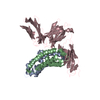


 PDBj
PDBj


 Assembly


 Type: D-saccharide, beta linking / Mass: 180.156 Da / Num. of mol.: 6
 Mass: 118.174 Da / Num. of mol.: 1 / Source method: obtained synthetically / Formula: C6H14O2 / Comment: precipitant*YM
 Mass: 18.015 Da / Num. of mol.: 55 / Source method: isolated from a natural source / Formula: H2O
 Sample preparation
 / Beamline: X10SA / Wavelength: 0.9079, 0.9795, 0.9797
 Processing