 Movie
Movie Controller

[English] 日本語
 Yorodumi
- PDB-6ih2: Crystal structure of Phosphite Dehydrogenase from Ralstonia sp. 4506 -
+ Open data

- Basic information
| Entry | Database: PDB / ID: 6ih2 | ||||||
|---|---|---|---|---|---|---|---|
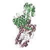
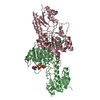
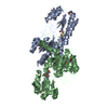
| Title | Crystal structure of Phosphite Dehydrogenase from Ralstonia sp. 4506 | ||||||
 Components Components | Phosphite dehydrogenase | ||||||
 Keywords Keywords |  OXIDOREDUCTASE / Phosphite Dehydrogenase OXIDOREDUCTASE / Phosphite Dehydrogenase | ||||||
| Function / homology |  Function and homology information Function and homology informationoxidoreductase activity, acting on the CH-OH group of donors, NAD or NADP as acceptor / NAD binding Similarity search - Function | ||||||
| Biological species |  Ralstonia sp. 4506 (bacteria) Ralstonia sp. 4506 (bacteria) | ||||||
| Method | X-RAY DIFFRACTION / SYNCHROTRON / MOLECULAR REPLACEMENT / Resolution: 2.048 Å | ||||||
 Authors Authors | Song, X. / Zhao, Z. / Liu, Y. / Feng, Y. | ||||||
 Citation Citation | Journal: Acs Catalysis / Year: 2019 Title: Structural Insights into Phosphite Dehydrogenase Variants Favoring a Non-natural Redox Cofactor Authors: Liu, Y. / Feng, Y. / Wang, L. / Guo, X. / Liu, W. / Li, Q. / Wang, X. / Xue, S. / Zhao, Z. | ||||||
| History |
|
- Structure visualization
| Structure viewer | Molecule: MolmilJmol/JSmol |
|---|
- Downloads & links
-Download
| PDBx/mmCIF format | 6ih2.cif.gz | 148.7 KB | Display | PDBx/mmCIF format |
|---|---|---|---|---|
| PDB format | pdb6ih2.ent.gz | 115.6 KB | Display | PDB format |
| PDBx/mmJSON format | 6ih2.json.gz | Tree view | PDBx/mmJSON format | |
| Others |  Other downloads Other downloads |
-Validation report
| Arichive directory | https://data.pdbj.org/pub/pdb/validation_reports/ih/6ih2ftp://data.pdbj.org/pub/pdb/validation_reports/ih/6ih2 | HTTPS FTP |
|---|
-Related structure data
| Related structure data |  6ih3C  6ih4C  6ih5C  6ih6C 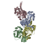 6ih8C 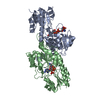 4e5mS S: Starting model for refinement C: citing same article ( |
|---|---|
| Similar structure data |


-Links
 PDBj
PDBj- Assembly
| Deposited unit | 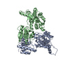
| ||||||||
|---|---|---|---|---|---|---|---|---|---|
| 1 | 
| ||||||||
| Unit cell |
|
-Components
| #1: Protein | Mass: 36627.430 Da / Num. of mol.: 2 Source method: isolated from a genetically manipulated source Source: (gene. exp.) Ralstonia sp. 4506 (bacteria) / Gene: ptxD / Production host: Escherichia coli (E. coli) / References: UniProt: G4XDR8#2: Water | ChemComp-HOH / | Water Mass: 18.015 Da / Num. of mol.: 567 / Source method: isolated from a natural source / Formula: H2O Mass: 18.015 Da / Num. of mol.: 567 / Source method: isolated from a natural source / Formula: H2O |
|---|
-Experimental details
-Experiment
| Experiment | Method: X-RAY DIFFRACTION / Number of used crystals: 1 |
|---|
- Sample preparation
| Crystal | Density Matthews: 2.52 Å3/Da / Density % sol: 51.21 % |
|---|---|
| Crystal grow | Temperature: 289 K / Method: evaporation / pH: 8 Details: 0.2 M NaCl, 0.1 M Tris-HCl pH 8.0, 25% (w/v) PEG3350 |
-Data collection
| Diffraction | Mean temperature: 100 K / Serial crystal experiment: N |
|---|---|
| Diffraction source | Source: SYNCHROTRON / Site: SSRF  / Beamline: BL18U1 / Wavelength: 0.9793 Å / Beamline: BL18U1 / Wavelength: 0.9793 Å |
| Detector | Type: MAR CCD 165 mm / Detector: CCD / Date: Dec 9, 2017 |
| Radiation | Protocol: SINGLE WAVELENGTH / Monochromatic (M) / Laue (L): M / Scattering type: x-ray |
| Radiation wavelength | Wavelength: 0.9793 Å / Relative weight: 1 |
| Reflection | Resolution: 2.048→38.324 Å / Num. obs: 46894 / % possible obs: 98.79 % / Redundancy: 12.3 % / Rmerge(I) obs: 0.13 / Net I/σ(I): 6.6 |
| Reflection shell | Resolution: 2.05→2.12 Å / Redundancy: 12.2 % / Rmerge(I) obs: 0.46 / Num. unique obs: 4180 / % possible all: 89.79 |
- Processing
| Software |
| ||||||||||||||||||||||||||||||||||||||||||||||||||||||||||||||||||||||||||||||||||||||||||||||||||||||||||||||||||||||||||||||
|---|---|---|---|---|---|---|---|---|---|---|---|---|---|---|---|---|---|---|---|---|---|---|---|---|---|---|---|---|---|---|---|---|---|---|---|---|---|---|---|---|---|---|---|---|---|---|---|---|---|---|---|---|---|---|---|---|---|---|---|---|---|---|---|---|---|---|---|---|---|---|---|---|---|---|---|---|---|---|---|---|---|---|---|---|---|---|---|---|---|---|---|---|---|---|---|---|---|---|---|---|---|---|---|---|---|---|---|---|---|---|---|---|---|---|---|---|---|---|---|---|---|---|---|---|---|---|---|
| Refinement | Method to determine structure: MOLECULAR REPLACEMENT Starting model: 4E5M Resolution: 2.048→38.324 Å / SU ML: 0.16 / Cross valid method: FREE R-VALUE / σ(F): 1.36 / Phase error: 18.06 / Stereochemistry target values: ML
| ||||||||||||||||||||||||||||||||||||||||||||||||||||||||||||||||||||||||||||||||||||||||||||||||||||||||||||||||||||||||||||||
| Solvent computation | Shrinkage radii: 0.9 Å / VDW probe radii: 1.11 Å / Solvent model: FLAT BULK SOLVENT MODEL | ||||||||||||||||||||||||||||||||||||||||||||||||||||||||||||||||||||||||||||||||||||||||||||||||||||||||||||||||||||||||||||||
| Refinement step | Cycle: LAST / Resolution: 2.048→38.324 Å
| ||||||||||||||||||||||||||||||||||||||||||||||||||||||||||||||||||||||||||||||||||||||||||||||||||||||||||||||||||||||||||||||
| Refine LS restraints |
| ||||||||||||||||||||||||||||||||||||||||||||||||||||||||||||||||||||||||||||||||||||||||||||||||||||||||||||||||||||||||||||||
| LS refinement shell |
|
