 Movie
Movie Controller

[English] 日本語
 Yorodumi
- PDB-3qj9: Crystal structure of fatty acid amide hydrolase with small molecu... -
+ Open data

- Basic information
| Entry | Database: PDB / ID: 3qj9 | ||||||
|---|---|---|---|---|---|---|---|
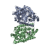
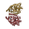
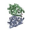
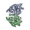
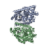
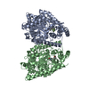
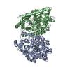
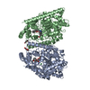
| Title | Crystal structure of fatty acid amide hydrolase with small molecule inhibitor | ||||||
 Components Components | Fatty-acid amide hydrolase 1 | ||||||
 Keywords Keywords | HYDROLASE/HYDROLASE INHIBITOR / protein-inhibitor complex / FAAH / Fatty-acid amide hydrolase / HYDROLASE-HYDROLASE INHIBITOR complex | ||||||
| Function / homology |  Function and homology information Function and homology information Arachidonic acid metabolism / fatty acid amide hydrolase / fatty acid amide hydrolase activity / monoacylglycerol catabolic process / acylglycerol lipase activity / amidase activity / fatty acid catabolic process / Hydrolases; Acting on ester bonds; Carboxylic-ester hydrolases / hydrolase activity, acting on ester bonds / organelle membrane ...Arachidonic acid metabolism / fatty acid amide hydrolase / fatty acid amide hydrolase activity / monoacylglycerol catabolic process / acylglycerol lipase activity / amidase activity / fatty acid catabolic process / Hydrolases; Acting on ester bonds; Carboxylic-ester hydrolases / hydrolase activity, acting on ester bonds / organelle membrane / positive regulation of vasoconstriction / fatty acid metabolic process / phospholipid binding / Golgi membrane / lipid binding / endoplasmic reticulum membrane / identical protein binding Arachidonic acid metabolism / fatty acid amide hydrolase / fatty acid amide hydrolase activity / monoacylglycerol catabolic process / acylglycerol lipase activity / amidase activity / fatty acid catabolic process / Hydrolases; Acting on ester bonds; Carboxylic-ester hydrolases / hydrolase activity, acting on ester bonds / organelle membrane ...Arachidonic acid metabolism / fatty acid amide hydrolase / fatty acid amide hydrolase activity / monoacylglycerol catabolic process / acylglycerol lipase activity / amidase activity / fatty acid catabolic process / Hydrolases; Acting on ester bonds; Carboxylic-ester hydrolases / hydrolase activity, acting on ester bonds / organelle membrane / positive regulation of vasoconstriction / fatty acid metabolic process / phospholipid binding / Golgi membrane / lipid binding / endoplasmic reticulum membrane / identical protein bindingSimilarity search - Function | ||||||
| Biological species |  Rattus norvegicus (Norway rat) Rattus norvegicus (Norway rat) | ||||||
| Method | X-RAY DIFFRACTION / SYNCHROTRON / MOLECULAR REPLACEMENT / Resolution: 2.3 Å | ||||||
 Authors Authors | Min, X. / Walker, N.P.C. / Wang, Z. | ||||||
 Citation Citation | Journal: Proc.Natl.Acad.Sci.USA / Year: 2011 Title: Discovery and molecular basis of potent noncovalent inhibitors of fatty acid amide hydrolase (FAAH). Authors: Min, X. / Thibault, S.T. / Porter, A.C. / Gustin, D.J. / Carlson, T.J. / Xu, H. / Lindstrom, M. / Xu, G. / Uyeda, C. / Ma, Z. / Li, Y. / Kayser, F. / Walker, N.P. / Wang, Z. | ||||||
| History |
|
- Structure visualization
| Structure viewer | Molecule: MolmilJmol/JSmol |
|---|
- Downloads & links
-Download
| PDBx/mmCIF format | 3qj9.cif.gz | 239.6 KB | Display | PDBx/mmCIF format |
|---|---|---|---|---|
| PDB format | pdb3qj9.ent.gz | 188.4 KB | Display | PDB format |
| PDBx/mmJSON format | 3qj9.json.gz | Tree view | PDBx/mmJSON format | |
| Others |  Other downloads Other downloads |
-Validation report
| Arichive directory | https://data.pdbj.org/pub/pdb/validation_reports/qj/3qj9ftp://data.pdbj.org/pub/pdb/validation_reports/qj/3qj9 | HTTPS FTP |
|---|
-Related structure data


-Links
 PDBj
PDBj- Assembly
| Deposited unit | 
| ||||||||
|---|---|---|---|---|---|---|---|---|---|
| 1 |
| ||||||||
| Unit cell |
|
-Components
-Protein , 1 types, 2 molecules AB
| #1: Protein | Mass: 64593.180 Da / Num. of mol.: 2 / Fragment: Residues 32-579 Source method: isolated from a genetically manipulated source Source: (gene. exp.) Rattus norvegicus (Norway rat) / Gene: Faah, Faah1 / Plasmid: pTrcHisA / Production host:  Escherichia coli (E. coli) / Strain (production host): BL21 / References: UniProt: P97612, fatty acid amide hydrolase Escherichia coli (E. coli) / Strain (production host): BL21 / References: UniProt: P97612, fatty acid amide hydrolase |
|---|
-Non-polymers , 5 types, 722 molecules 


| #2: Chemical |  Mass: 439.509 Da / Num. of mol.: 2 / Source method: obtained synthetically / Formula: C26H25N5O2 Mass: 439.509 Da / Num. of mol.: 2 / Source method: obtained synthetically / Formula: C26H25N5O2#3: Chemical | ChemComp-EDO / Ethylene glycol Mass: 62.068 Da / Num. of mol.: 21 / Source method: obtained synthetically / Formula: C2H6O2 Mass: 62.068 Da / Num. of mol.: 21 / Source method: obtained synthetically / Formula: C2H6O2#4: Chemical | Glycerol Mass: 92.094 Da / Num. of mol.: 2 / Source method: obtained synthetically / Formula: C3H8O3 Mass: 92.094 Da / Num. of mol.: 2 / Source method: obtained synthetically / Formula: C3H8O3#5: Chemical | ChemComp-PGE / | Polyethylene glycol Mass: 150.173 Da / Num. of mol.: 1 / Source method: obtained synthetically / Formula: C6H14O4 Mass: 150.173 Da / Num. of mol.: 1 / Source method: obtained synthetically / Formula: C6H14O4#6: Water | ChemComp-HOH / | WaterMass: 18.015 Da / Num. of mol.: 696 / Source method: isolated from a natural source / Formula: H2O |
|---|
-Experimental details
-Experiment
| Experiment | Method: X-RAY DIFFRACTION / Number of used crystals: 1 |
|---|
- Sample preparation
| Crystal | Density Matthews: 2.76 Å3/Da / Density % sol: 55.38 % |
|---|---|
| Crystal grow | Temperature: 289 K / Method: vapor diffusion, hanging drop / pH: 5.5 Details: PEG3350, NH4F, pH 5.5, vapor diffusion, hanging drop, temperature 289K |
-Data collection
| Diffraction | Mean temperature: 100 K | ||||||||||||||||||||||||||||||||||||||||||||||||||||||||||||||||||||||||||||||||||||||||
|---|---|---|---|---|---|---|---|---|---|---|---|---|---|---|---|---|---|---|---|---|---|---|---|---|---|---|---|---|---|---|---|---|---|---|---|---|---|---|---|---|---|---|---|---|---|---|---|---|---|---|---|---|---|---|---|---|---|---|---|---|---|---|---|---|---|---|---|---|---|---|---|---|---|---|---|---|---|---|---|---|---|---|---|---|---|---|---|---|---|
| Diffraction source | Source: SYNCHROTRON / Site: ALS  / Beamline: 5.0.2 / Wavelength: 1 / Wavelength: 1 Å / Beamline: 5.0.2 / Wavelength: 1 / Wavelength: 1 Å | ||||||||||||||||||||||||||||||||||||||||||||||||||||||||||||||||||||||||||||||||||||||||
| Detector | Type: ADSC QUANTUM 315 / Detector: CCD / Details: mirrors | ||||||||||||||||||||||||||||||||||||||||||||||||||||||||||||||||||||||||||||||||||||||||
| Radiation | Monochromator: Double-crystal, Si(111) / Protocol: SINGLE WAVELENGTH / Scattering type: x-ray | ||||||||||||||||||||||||||||||||||||||||||||||||||||||||||||||||||||||||||||||||||||||||
| Radiation wavelength | Wavelength: 1 Å / Relative weight: 1 | ||||||||||||||||||||||||||||||||||||||||||||||||||||||||||||||||||||||||||||||||||||||||
| Reflection | Resolution: 2.3→49.383 Å / Num. all: 60454 / Num. obs: 60454 / % possible obs: 95 % / Redundancy: 5.2 % / Rsym value: 0.148 / Net I/σ(I): 9.2 | ||||||||||||||||||||||||||||||||||||||||||||||||||||||||||||||||||||||||||||||||||||||||
| Reflection shell | Diffraction-ID: 1
|
- Processing
| Software |
| |||||||||||||||||||||||||||||||||||||||||||||||||||||||||||||||||
|---|---|---|---|---|---|---|---|---|---|---|---|---|---|---|---|---|---|---|---|---|---|---|---|---|---|---|---|---|---|---|---|---|---|---|---|---|---|---|---|---|---|---|---|---|---|---|---|---|---|---|---|---|---|---|---|---|---|---|---|---|---|---|---|---|---|---|
| Refinement | Method to determine structure: MOLECULAR REPLACEMENT / Resolution: 2.3→30 Å / Cor.coef. Fo:Fc: 0.938 / Cor.coef. Fo:Fc free: 0.908 / WRfactor Rfree: 0.2034 / WRfactor Rwork: 0.1668 / Occupancy max: 1 / Occupancy min: 0.5 / FOM work R set: 0.8827 / SU B: 5.222 / SU ML: 0.128 / SU R Cruickshank DPI: 0.2915 / SU Rfree: 0.213 / Cross valid method: THROUGHOUT / σ(F): 0 / ESU R Free: 0.213 / Stereochemistry target values: MAXIMUM LIKELIHOOD Details: HYDROGENS HAVE BEEN ADDED IN THE RIDING POSITIONS U VALUES : REFINED INDIVIDUALLY
| |||||||||||||||||||||||||||||||||||||||||||||||||||||||||||||||||
| Solvent computation | Ion probe radii: 0.8 Å / Shrinkage radii: 0.8 Å / VDW probe radii: 1.2 Å / Solvent model: MASK | |||||||||||||||||||||||||||||||||||||||||||||||||||||||||||||||||
| Displacement parameters | Biso max: 104.03 Å2 / Biso mean: 17.1351 Å2 / Biso min: 2 Å2
| |||||||||||||||||||||||||||||||||||||||||||||||||||||||||||||||||
| Refinement step | Cycle: LAST / Resolution: 2.3→30 Å
| |||||||||||||||||||||||||||||||||||||||||||||||||||||||||||||||||
| Refine LS restraints |
| |||||||||||||||||||||||||||||||||||||||||||||||||||||||||||||||||
| LS refinement shell | Resolution: 2.3→2.359 Å / Total num. of bins used: 20
|
