 Movie
Movie Controller

[English] 日本語
 Yorodumi
- PDB-3qgi: Crystal structure of the hepatitis C virus NS5B RNA-dependent RNA... -
+ Open data

- Basic information
| Entry | Database: PDB / ID: 3qgi | ||||||
|---|---|---|---|---|---|---|---|
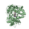
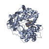
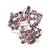
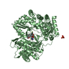
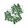
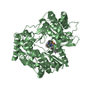
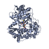
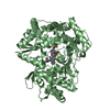
| Title | Crystal structure of the hepatitis C virus NS5B RNA-dependent RNA polymerase genotype 1a complex with N-[(2S)-butan-2-yl]-6-[(3R)-3-{[4-(trifluoromethoxy)benzyl]carbamoyl}-4-{[4-(trifluoromethoxy)phenyl]sulfonyl}piperazin-1-yl]pyridazine-3-carboxamide | ||||||
 Components Components | RNA-directed RNA polymerase RNA-dependent RNA polymerase RNA-dependent RNA polymerase | ||||||
 Keywords Keywords | TRANSFERASE/TRANSFERASE INHIBITOR / NS5B / POLYMERASE / HCV / FINGERS / PALM / THUMB / TRANSFERASE-TRANSFERASE INHIBITOR complex | ||||||
| Function / homology |  Function and homology informationhepacivirin / host cell mitochondrial membrane / host cell lipid droplet / symbiont-mediated suppression of host TRAF-mediated signal transduction / transformation of host cell by virus / symbiont-mediated perturbation of host cell cycle G1/S transition checkpoint / symbiont-mediated suppression of host JAK-STAT cascade via inhibition of STAT1 activity / symbiont-mediated suppression of host cytoplasmic pattern recognition receptor signaling pathway via inhibition of MAVS activity / SH3 domain binding / nucleoside-triphosphate phosphatase ...hepacivirin / host cell mitochondrial membrane / host cell lipid droplet / symbiont-mediated suppression of host TRAF-mediated signal transduction / transformation of host cell by virus / symbiont-mediated perturbation of host cell cycle G1/S transition checkpoint / symbiont-mediated suppression of host JAK-STAT cascade via inhibition of STAT1 activity / symbiont-mediated suppression of host cytoplasmic pattern recognition receptor signaling pathway via inhibition of MAVS activity / SH3 domain binding / nucleoside-triphosphate phosphatase / protein complex oligomerization / monoatomic ion channel activity / viral nucleocapsid / clathrin-dependent endocytosis of virus by host cell / host cell cytoplasm / Hydrolases; Acting on peptide bonds (peptidases); Cysteine endopeptidases / RNA helicase activity / host cell perinuclear region of cytoplasm / host cell endoplasmic reticulum membrane / symbiont-mediated suppression of host type I interferon-mediated signaling pathway / RNA helicase / ribonucleoprotein complex / induction by virus of host autophagy / RNA-directed RNA polymerase / virus-mediated perturbation of host defense response / viral RNA genome replication / cysteine-type endopeptidase activity / RNA-dependent RNA polymerase activity / serine-type endopeptidase activity / nucleotide binding / fusion of virus membrane with host endosome membrane / viral envelope / host cell nucleus / virion attachment to host cell / host cell plasma membrane / virion membrane / structural molecule activity / ATP hydrolysis activity / proteolysis / RNA binding / zinc ion binding / ATP binding / membrane Function and homology informationhepacivirin / host cell mitochondrial membrane / host cell lipid droplet / symbiont-mediated suppression of host TRAF-mediated signal transduction / transformation of host cell by virus / symbiont-mediated perturbation of host cell cycle G1/S transition checkpoint / symbiont-mediated suppression of host JAK-STAT cascade via inhibition of STAT1 activity / symbiont-mediated suppression of host cytoplasmic pattern recognition receptor signaling pathway via inhibition of MAVS activity / SH3 domain binding / nucleoside-triphosphate phosphatase ...hepacivirin / host cell mitochondrial membrane / host cell lipid droplet / symbiont-mediated suppression of host TRAF-mediated signal transduction / transformation of host cell by virus / symbiont-mediated perturbation of host cell cycle G1/S transition checkpoint / symbiont-mediated suppression of host JAK-STAT cascade via inhibition of STAT1 activity / symbiont-mediated suppression of host cytoplasmic pattern recognition receptor signaling pathway via inhibition of MAVS activity / SH3 domain binding / nucleoside-triphosphate phosphatase / protein complex oligomerization / monoatomic ion channel activity / viral nucleocapsid / clathrin-dependent endocytosis of virus by host cell / host cell cytoplasm / Hydrolases; Acting on peptide bonds (peptidases); Cysteine endopeptidases / RNA helicase activity / host cell perinuclear region of cytoplasm / host cell endoplasmic reticulum membrane / symbiont-mediated suppression of host type I interferon-mediated signaling pathway / RNA helicase / ribonucleoprotein complex / induction by virus of host autophagy / RNA-directed RNA polymerase / virus-mediated perturbation of host defense response / viral RNA genome replication / cysteine-type endopeptidase activity / RNA-dependent RNA polymerase activity / serine-type endopeptidase activity / nucleotide binding / fusion of virus membrane with host endosome membrane / viral envelope / host cell nucleus / virion attachment to host cell / host cell plasma membrane / virion membrane / structural molecule activity / ATP hydrolysis activity / proteolysis / RNA binding / zinc ion binding / ATP binding / membraneSimilarity search - Function | ||||||
| Biological species |  Hepatitis C virus subtype 1a Hepatitis C virus subtype 1a | ||||||
| Method | X-RAY DIFFRACTION / SYNCHROTRON / MOLECULAR REPLACEMENT / Resolution: 1.8 Å | ||||||
 Authors Authors | Sheriff, S. | ||||||
 Citation Citation | Journal: Bioorg.Med.Chem.Lett. / Year: 2011 Title: Investigation of the mode of binding of a novel series of N-benzyl-4-heteroaryl-1-(phenylsulfonyl)piperazine-2-carboxamides to the hepatitis C virus polymerase. Authors: Gentles, R.G. / Sheriff, S. / Beno, B.R. / Wan, C. / Kish, K. / Ding, M. / Zheng, X. / Chupak, L. / Poss, M.A. / Witmer, M.R. / Morin, P. / Wang, Y.K. / Rigat, K. / Lemm, J. / Voss, S. / ...Authors: Gentles, R.G. / Sheriff, S. / Beno, B.R. / Wan, C. / Kish, K. / Ding, M. / Zheng, X. / Chupak, L. / Poss, M.A. / Witmer, M.R. / Morin, P. / Wang, Y.K. / Rigat, K. / Lemm, J. / Voss, S. / Liu, M. / Pelosi, L. / Roberts, S.B. / Gao, M. / Kadow, J.F. | ||||||
| History |
|
- Structure visualization
| Structure viewer | Molecule: MolmilJmol/JSmol |
|---|
- Downloads & links
-Download
| PDBx/mmCIF format | 3qgi.cif.gz | 138 KB | Display | PDBx/mmCIF format |
|---|---|---|---|---|
| PDB format | pdb3qgi.ent.gz | 105.3 KB | Display | PDB format |
| PDBx/mmJSON format | 3qgi.json.gz | Tree view | PDBx/mmJSON format | |
| Others |  Other downloads Other downloads |
-Validation report
| Arichive directory | https://data.pdbj.org/pub/pdb/validation_reports/qg/3qgiftp://data.pdbj.org/pub/pdb/validation_reports/qg/3qgi | HTTPS FTP |
|---|
-Related structure data
| Related structure data |  3qgdC  3qgeC  3qgfC  3qggC  3qghC  3xxxS C: citing same article ( S: Starting model for refinement |
|---|---|
| Similar structure data |


-Links
 PDBj
PDBj
- Assembly
| Deposited unit | 
| ||||||||
|---|---|---|---|---|---|---|---|---|---|
| 1 |
| ||||||||
| Unit cell |
|
-Components
| #1: Protein | RNA-dependent RNA polymerase Mass: 63018.320 Da / Num. of mol.: 1 Source method: isolated from a genetically manipulated source Source: (gene. exp.) Hepatitis C virus subtype 1a / Strain: Bartenschlager / Gene: NS5B / Plasmid: pET21b / Production host:  Escherichia coli (E. coli) / Strain (production host): ROSETTA2 (DE3) Escherichia coli (E. coli) / Strain (production host): ROSETTA2 (DE3)References: UniProt: B1PBP5, UniProt: P26664*PLUS, RNA-directed RNA polymerase | ||||
|---|---|---|---|---|---|
| #2: Chemical | ChemComp-33F /   Mass: 704.641 Da / Num. of mol.: 1 / Source method: obtained synthetically / Formula: C29H30F6N6O6S Mass: 704.641 Da / Num. of mol.: 1 / Source method: obtained synthetically / Formula: C29H30F6N6O6S | ||||
| #3: Chemical | ChemComp-GOL / Glycerol  Mass: 92.094 Da / Num. of mol.: 14 / Source method: obtained synthetically / Formula: C3H8O3 Mass: 92.094 Da / Num. of mol.: 14 / Source method: obtained synthetically / Formula: C3H8O3#4: Water | ChemComp-HOH / | Water Mass: 18.015 Da / Num. of mol.: 541 / Source method: isolated from a natural source / Formula: H2O Mass: 18.015 Da / Num. of mol.: 541 / Source method: isolated from a natural source / Formula: H2OSequence details | THE UNIPROT ENTRY B1PBP5_9HEPC DOES NOT PROVIDE THE COMPLETE SEQUENCE. THE COMPLETE SEQUENCE CAN BE ...THE UNIPROT ENTRY B1PBP5_9HEPC DOES NOT PROVIDE THE COMPLETE SEQUENCE. THE COMPLETE SEQUENCE CAN BE FOUND IN THE NCBI ENTRY NP_751928.1. | |
-Experimental details
-Experiment
| Experiment | Method: X-RAY DIFFRACTION / Number of used crystals: 1 |
|---|
- Sample preparation
| Crystal | Density Matthews: 2.72 Å3/Da / Density % sol: 54.72 % |
|---|---|
| Crystal grow | Temperature: 277 K / Method: vapor diffusion, hanging drop / pH: 7.3 Details: 25.5% (w/v) PEG 4000, 170 mM (NH4)2SO4, 15% (v/v) glycerol, pH 7.3, vapor diffusion, hanging drop, temperature 277K |
-Data collection
| Diffraction | Mean temperature: 100 K |
|---|---|
| Diffraction source | Source: SYNCHROTRON / Site: APS  / Beamline: 17-BM / Wavelength: 1 / Wavelength: 1 Å / Beamline: 17-BM / Wavelength: 1 / Wavelength: 1 Å |
| Detector | Type: MAR CCD 165 mm / Detector: CCD / Date: Mar 29, 2008 |
| Radiation | Protocol: SINGLE WAVELENGTH / Monochromatic (M) / Laue (L): M / Scattering type: x-ray |
| Radiation wavelength | Wavelength: 1 Å / Relative weight: 1 |
| Reflection | Resolution: 1.8→50 Å / Num. obs: 62602 / % possible obs: 97.6 % / Observed criterion σ(I): 0 / Redundancy: 4.8 % / Biso Wilson estimate: 16.6 Å2 / Rmerge(I) obs: 0.089 / Net I/σ(I): 18.2 |
| Reflection shell | Resolution: 1.8→1.86 Å / Redundancy: 4.9 % / Rmerge(I) obs: 0.46 / Mean I/σ(I) obs: 3.5 / % possible all: 96.2 |
- Processing
| Software |
| ||||||||||||||||||||||||||||||||||||||||||||||||||||||||||||||||||||||||||||||||||||||||||||||||||||||||||||
|---|---|---|---|---|---|---|---|---|---|---|---|---|---|---|---|---|---|---|---|---|---|---|---|---|---|---|---|---|---|---|---|---|---|---|---|---|---|---|---|---|---|---|---|---|---|---|---|---|---|---|---|---|---|---|---|---|---|---|---|---|---|---|---|---|---|---|---|---|---|---|---|---|---|---|---|---|---|---|---|---|---|---|---|---|---|---|---|---|---|---|---|---|---|---|---|---|---|---|---|---|---|---|---|---|---|---|---|---|---|
| Refinement | Method to determine structure: MOLECULAR REPLACEMENT Starting model: PDB ENTRY 3XXX, NS5B 1A Resolution: 1.8→26.35 Å / Cor.coef. Fo:Fc: 0.9515 / Cor.coef. Fo:Fc free: 0.9362 / Occupancy max: 1 / Occupancy min: 0.5 / SU R Cruickshank DPI: 0.1 / Cross valid method: THROUGHOUT / σ(F): 0 / SU R Blow DPI: 0.107 / SU Rfree Blow DPI: 0.1 / SU Rfree Cruickshank DPI: 0.097
| ||||||||||||||||||||||||||||||||||||||||||||||||||||||||||||||||||||||||||||||||||||||||||||||||||||||||||||
| Displacement parameters | Biso max: 106.13 Å2 / Biso mean: 16.9397 Å2 / Biso min: 4.24 Å2
| ||||||||||||||||||||||||||||||||||||||||||||||||||||||||||||||||||||||||||||||||||||||||||||||||||||||||||||
| Refine analyze | Luzzati coordinate error obs: 0.162 Å | ||||||||||||||||||||||||||||||||||||||||||||||||||||||||||||||||||||||||||||||||||||||||||||||||||||||||||||
| Refinement step | Cycle: LAST / Resolution: 1.8→26.35 Å
| ||||||||||||||||||||||||||||||||||||||||||||||||||||||||||||||||||||||||||||||||||||||||||||||||||||||||||||
| Refine LS restraints |
| ||||||||||||||||||||||||||||||||||||||||||||||||||||||||||||||||||||||||||||||||||||||||||||||||||||||||||||
| LS refinement shell | Resolution: 1.8→1.85 Å / Total num. of bins used: 20
|
