 Movie
Movie Controller

[English] 日本語
 Yorodumi
- PDB-3qgf: Crystal structure of the hepatitis C virus NS5B RNA-dependent RNA... -
+ Open data

- Basic information
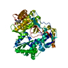
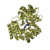
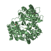
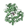
| Entry | Database: PDB / ID: 3qgf | ||||||
|---|---|---|---|---|---|---|---|
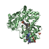
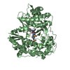
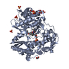
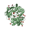
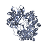
| Title | Crystal structure of the hepatitis C virus NS5B RNA-dependent RNA polymerase complex with (2E)-3-(4-{[(1-{[(13-cyclohexyl-6-oxo-6,7-dihydro-5H-indolo[1,2-d][1,4]benzodiazepin-10-yl)carbonyl]amino}cyclopentyl)carbonyl]amino}phenyl)prop-2-enoic acid and (2R)-4-(6-chloropyridazin-3-yl)-N-(4-methoxybenzyl)-1-{[4-(trifluoromethoxy)phenyl]sulfonyl}piperazine-2-carboxamide | ||||||
 Components Components | RNA-directed RNA polymerase RNA-dependent RNA polymerase RNA-dependent RNA polymerase | ||||||
 Keywords Keywords | TRANSFERASE/TRANSFERASE INHIBITOR / NS5B / POLYMERASE / HCV / FINGERS / PALM / THUMB / TRANSFERASE-TRANSFERASE INHIBITOR complex | ||||||
| Function / homology |  Function and homology information Function and homology informationpositive stranded viral RNA replication / hepacivirin / host cell mitochondrial membrane / host cell lipid droplet / symbiont-mediated suppression of host TRAF-mediated signal transduction / transformation of host cell by virus / symbiont-mediated perturbation of host cell cycle G1/S transition checkpoint / symbiont-mediated suppression of host JAK-STAT cascade via inhibition of STAT1 activity / symbiont-mediated suppression of host cytoplasmic pattern recognition receptor signaling pathway via inhibition of MAVS activity / viral genome replication ...positive stranded viral RNA replication / hepacivirin / host cell mitochondrial membrane / host cell lipid droplet / symbiont-mediated suppression of host TRAF-mediated signal transduction / transformation of host cell by virus / symbiont-mediated perturbation of host cell cycle G1/S transition checkpoint / symbiont-mediated suppression of host JAK-STAT cascade via inhibition of STAT1 activity / symbiont-mediated suppression of host cytoplasmic pattern recognition receptor signaling pathway via inhibition of MAVS activity / viral genome replication / SH3 domain binding / : / nucleoside-triphosphate phosphatase / protein complex oligomerization / monoatomic ion channel activity / viral nucleocapsid / clathrin-dependent endocytosis of virus by host cell / Hydrolases; Acting on peptide bonds (peptidases); Cysteine endopeptidases / RNA helicase activity / host cell endoplasmic reticulum membrane / host cell perinuclear region of cytoplasm / RNA helicase / induction by virus of host autophagy / ribonucleoprotein complex / RNA-directed RNA polymerase / viral RNA genome replication / cysteine-type endopeptidase activity / RNA-dependent RNA polymerase activity / serine-type endopeptidase activity / fusion of virus membrane with host endosome membrane / viral envelope / symbiont-mediated suppression of host type I interferon-mediated signaling pathway / host cell nucleus / virion attachment to host cell / host cell plasma membrane / virion membrane / structural molecule activity / ATP hydrolysis activity / protein-containing complex / proteolysis / RNA binding / zinc ion binding / ATP binding / membrane / identical protein bindingSimilarity search - Function | ||||||
| Biological species |  Hepatitis C virus subtype 1b Hepatitis C virus subtype 1b | ||||||
| Method | X-RAY DIFFRACTION / SYNCHROTRON / MOLECULAR REPLACEMENT / Resolution: 2.45 Å | ||||||
 Authors Authors | Sheriff, S. | ||||||
 Citation Citation | Journal: Bioorg.Med.Chem.Lett. / Year: 2011 Title: Investigation of the mode of binding of a novel series of N-benzyl-4-heteroaryl-1-(phenylsulfonyl)piperazine-2-carboxamides to the hepatitis C virus polymerase. Authors: Gentles, R.G. / Sheriff, S. / Beno, B.R. / Wan, C. / Kish, K. / Ding, M. / Zheng, X. / Chupak, L. / Poss, M.A. / Witmer, M.R. / Morin, P. / Wang, Y.K. / Rigat, K. / Lemm, J. / Voss, S. / ...Authors: Gentles, R.G. / Sheriff, S. / Beno, B.R. / Wan, C. / Kish, K. / Ding, M. / Zheng, X. / Chupak, L. / Poss, M.A. / Witmer, M.R. / Morin, P. / Wang, Y.K. / Rigat, K. / Lemm, J. / Voss, S. / Liu, M. / Pelosi, L. / Roberts, S.B. / Gao, M. / Kadow, J.F. | ||||||
| History |
|
- Structure visualization
| Structure viewer | Molecule: MolmilJmol/JSmol |
|---|
- Downloads & links
-Download
| PDBx/mmCIF format | 3qgf.cif.gz | 228.2 KB | Display | PDBx/mmCIF format |
|---|---|---|---|---|
| PDB format | pdb3qgf.ent.gz | 181.9 KB | Display | PDB format |
| PDBx/mmJSON format | 3qgf.json.gz | Tree view | PDBx/mmJSON format | |
| Others |  Other downloads Other downloads |
-Validation report
| Arichive directory | https://data.pdbj.org/pub/pdb/validation_reports/qg/3qgfftp://data.pdbj.org/pub/pdb/validation_reports/qg/3qgf | HTTPS FTP |
|---|
-Related structure data
| Related structure data |  3qgdC  3qgeC  3qggC  3qghC  3qgiC  3q0zS C: citing same article ( S: Starting model for refinement |
|---|---|
| Similar structure data |


-Links
 PDBj
PDBj
- Assembly
| Deposited unit | 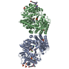
| ||||||||
|---|---|---|---|---|---|---|---|---|---|
| 1 | 
| ||||||||
| 2 | 
| ||||||||
| Unit cell |
|
-Components
| #1: Protein | RNA-dependent RNA polymerase / NS5B / p68 Mass: 63961.516 Da / Num. of mol.: 2 Source method: isolated from a genetically manipulated source Source: (gene. exp.) Hepatitis C virus subtype 1b / Strain: Bartenschlager / Gene: NS5B / Plasmid: pET21b / Production host:  Escherichia coli (E. coli) / Strain (production host): BL21(DE3) STAR / References: UniProt: Q9WMX2, RNA-directed RNA polymerase Escherichia coli (E. coli) / Strain (production host): BL21(DE3) STAR / References: UniProt: Q9WMX2, RNA-directed RNA polymerase#2: Chemical |   Mass: 630.732 Da / Num. of mol.: 2 / Source method: obtained synthetically / Formula: C38H38N4O5 Mass: 630.732 Da / Num. of mol.: 2 / Source method: obtained synthetically / Formula: C38H38N4O5#3: Chemical |   Mass: 585.983 Da / Num. of mol.: 2 / Source method: obtained synthetically / Formula: C24H23ClF3N5O5S Mass: 585.983 Da / Num. of mol.: 2 / Source method: obtained synthetically / Formula: C24H23ClF3N5O5S#4: Chemical | ChemComp-SO4 / Sulfate  Mass: 96.063 Da / Num. of mol.: 10 / Source method: obtained synthetically / Formula: SO4 Mass: 96.063 Da / Num. of mol.: 10 / Source method: obtained synthetically / Formula: SO4#5: Water | ChemComp-HOH / | Water Mass: 18.015 Da / Num. of mol.: 252 / Source method: isolated from a natural source / Formula: H2O Mass: 18.015 Da / Num. of mol.: 252 / Source method: isolated from a natural source / Formula: H2O |
|---|
-Experimental details
-Experiment
| Experiment | Method: X-RAY DIFFRACTION / Number of used crystals: 1 |
|---|
- Sample preparation
| Crystal | Density Matthews: 3.05 Å3/Da / Density % sol: 59.68 % |
|---|---|
| Crystal grow | Temperature: 293 K / Method: vapor diffusion, hanging drop / pH: 5.6 Details: 100 mM Sodium Citrate, 1.75 M (NH4)2SO4, pH 5.6, vapor diffusion, hanging drop, temperature 293K |
-Data collection
| Diffraction | Mean temperature: 100 K |
|---|---|
| Diffraction source | Source: SYNCHROTRON / Site: APS  / Beamline: 17-ID / Wavelength: 1 Å / Beamline: 17-ID / Wavelength: 1 Å |
| Detector | Type: ADSC QUANTUM 210 / Detector: CCD / Date: Jun 28, 2007 |
| Radiation | Protocol: SINGLE WAVELENGTH / Monochromatic (M) / Laue (L): M / Scattering type: x-ray |
| Radiation wavelength | Wavelength: 1 Å / Relative weight: 1 |
| Reflection | Resolution: 2.45→50 Å / Num. obs: 50824 / % possible obs: 86 % / Observed criterion σ(I): 0 / Redundancy: 4.6 % / Biso Wilson estimate: 37.97 Å2 / Rmerge(I) obs: 0.093 / Net I/σ(I): 14.9 |
| Reflection shell | Resolution: 2.45→2.54 Å / Redundancy: 4.8 % / Rmerge(I) obs: 0.379 / Mean I/σ(I) obs: 4.3 / % possible all: 80.1 |
- Processing
| Software |
| ||||||||||||||||||||||||||||||||||||||||||||||||||||||||||||||||||||||||||||||||||||||||||||||||||||||||||||
|---|---|---|---|---|---|---|---|---|---|---|---|---|---|---|---|---|---|---|---|---|---|---|---|---|---|---|---|---|---|---|---|---|---|---|---|---|---|---|---|---|---|---|---|---|---|---|---|---|---|---|---|---|---|---|---|---|---|---|---|---|---|---|---|---|---|---|---|---|---|---|---|---|---|---|---|---|---|---|---|---|---|---|---|---|---|---|---|---|---|---|---|---|---|---|---|---|---|---|---|---|---|---|---|---|---|---|---|---|---|
| Refinement | Method to determine structure: MOLECULAR REPLACEMENT Starting model: PDB ENTRY 3Q0Z, NS5B Resolution: 2.45→35.44 Å / Cor.coef. Fo:Fc: 0.9139 / Cor.coef. Fo:Fc free: 0.8672 / Occupancy max: 1 / Occupancy min: 1 / SU R Cruickshank DPI: 0.447 / Cross valid method: THROUGHOUT / σ(F): 0 / SU R Blow DPI: 0.456 / SU Rfree Blow DPI: 0.289 / SU Rfree Cruickshank DPI: 0.291
| ||||||||||||||||||||||||||||||||||||||||||||||||||||||||||||||||||||||||||||||||||||||||||||||||||||||||||||
| Displacement parameters | Biso max: 189.5 Å2 / Biso mean: 33.8395 Å2 / Biso min: 4.72 Å2
| ||||||||||||||||||||||||||||||||||||||||||||||||||||||||||||||||||||||||||||||||||||||||||||||||||||||||||||
| Refine analyze | Luzzati coordinate error obs: 0.384 Å | ||||||||||||||||||||||||||||||||||||||||||||||||||||||||||||||||||||||||||||||||||||||||||||||||||||||||||||
| Refinement step | Cycle: LAST / Resolution: 2.45→35.44 Å
| ||||||||||||||||||||||||||||||||||||||||||||||||||||||||||||||||||||||||||||||||||||||||||||||||||||||||||||
| Refine LS restraints |
| ||||||||||||||||||||||||||||||||||||||||||||||||||||||||||||||||||||||||||||||||||||||||||||||||||||||||||||
| LS refinement shell | Resolution: 2.45→2.51 Å / Total num. of bins used: 20
|
