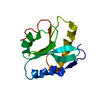
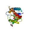
 Movie
Movie Controller


+ Open data

- Basic information
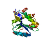
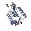
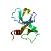
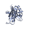
| Entry | Database: PDB / ID: 3q2b | ||||||
|---|---|---|---|---|---|---|---|
| Title | Crystal Structure of an Actin Depolymerizing Factor | ||||||
 Components Components | Cofilin/actin-depolymerizing factor homolog 1 | ||||||
 Keywords Keywords |  Actin-BINDING PROTEIN / Actin Regulator / Actin Actin-BINDING PROTEIN / Actin Regulator / Actin | ||||||
| Function / homology |  Function and homology information Function and homology informationcytoplasmic actin-based contraction involved in cell motility / actin filament depolymerization / actin monomer binding / actin cytoskeleton / actin cytoskeleton organization / cytoplasmSimilarity search - Function | ||||||
| Biological species |  Plasmodium falciparum (malaria parasite P. falciparum) Plasmodium falciparum (malaria parasite P. falciparum) | ||||||
| Method | X-RAY DIFFRACTION / SYNCHROTRON / MOLECULAR REPLACEMENT / Resolution: 1.6 Å | ||||||
 Authors Authors | Wong, W. / Clarke, O.B. / Gulbis, J.M. / Baum, J. | ||||||
 Citation Citation | Journal: Proc.Natl.Acad.Sci.USA / Year: 2011 Title: Minimal requirements for actin filament disassembly revealed by structural analysis of malaria parasite actin-depolymerizing factor 1 Authors: Wong, W. / Skau, C.T. / Marapana, D.S. / Hanssen, E. / Taylor, N.L. / Riglar, D.T. / Zuccala, E.S. / Angrisano, F. / Lewis, H. / Catimel, B. / Clarke, O.B. / Kershaw, N.J. / Perugini, M.A. / ...Authors: Wong, W. / Skau, C.T. / Marapana, D.S. / Hanssen, E. / Taylor, N.L. / Riglar, D.T. / Zuccala, E.S. / Angrisano, F. / Lewis, H. / Catimel, B. / Clarke, O.B. / Kershaw, N.J. / Perugini, M.A. / Kovar, D.R. / Gulbis, J.M. / Baum, J. | ||||||
| History |
|
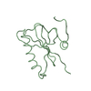
- Structure visualization
| Structure viewer | Molecule: MolmilJmol/JSmol |
|---|
- Downloads & links
-Download
| PDBx/mmCIF format | 3q2b.cif.gz | 67.7 KB | Display | PDBx/mmCIF format |
|---|---|---|---|---|
| PDB format | pdb3q2b.ent.gz | 49.7 KB | Display | PDB format |
| PDBx/mmJSON format | 3q2b.json.gz | Tree view | PDBx/mmJSON format | |
| Others |  Other downloads Other downloads |
-Validation report
| Arichive directory | https://data.pdbj.org/pub/pdb/validation_reports/q2/3q2bftp://data.pdbj.org/pub/pdb/validation_reports/q2/3q2b | HTTPS FTP |
|---|
-Related structure data
| Related structure data |  1f7sS S: Starting model for refinement |
|---|---|
| Similar structure data |
-Links
 PDBj
PDBj


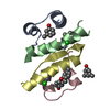
- Assembly
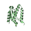
| Deposited unit | 
| ||||||||
|---|---|---|---|---|---|---|---|---|---|
| 1 |
| ||||||||
| Unit cell |
|
-Components
| #1: Protein | Mass: 13900.958 Da / Num. of mol.: 1 Source method: isolated from a genetically manipulated source Source: (gene. exp.) Plasmodium falciparum (malaria parasite P. falciparum)Strain: HB3 / Plasmid: pGEX4T / Production host:  Escherichia coli (E. coli) / Strain (production host): BL21(DE3) / References: UniProt: P86292 Escherichia coli (E. coli) / Strain (production host): BL21(DE3) / References: UniProt: P86292 | ||||
|---|---|---|---|---|---|
| #2: Chemical | 2-Mercaptoethanol  Mass: 78.133 Da / Num. of mol.: 2 / Source method: obtained synthetically / Formula: C2H6OS Mass: 78.133 Da / Num. of mol.: 2 / Source method: obtained synthetically / Formula: C2H6OS#3: Chemical | Tartaric acid  Mass: 150.087 Da / Num. of mol.: 2 / Source method: obtained synthetically / Formula: C4H6O6 Mass: 150.087 Da / Num. of mol.: 2 / Source method: obtained synthetically / Formula: C4H6O6#4: Water | ChemComp-HOH / | Water Mass: 18.015 Da / Num. of mol.: 77 / Source method: isolated from a natural source / Formula: H2O Mass: 18.015 Da / Num. of mol.: 77 / Source method: isolated from a natural source / Formula: H2O |
-Experimental details
-Experiment
| Experiment | Method: X-RAY DIFFRACTION / Number of used crystals: 1 |
|---|
- Sample preparation
| Crystal | Density Matthews: 1.94 Å3/Da / Density % sol: 36.75 % |
|---|---|
| Crystal grow | Temperature: 295 K / Method: vapor diffusion, hanging drop / pH: 4.6 Details: 1.8M (NH4)2SO4, 0.2M KNa tartrate, 0.1M NaOAc, pH 4.6, VAPOR DIFFUSION, HANGING DROP, temperature 295K |
-Data collection
| Diffraction source | Source: SYNCHROTRON / Site: Australian Synchrotron  / Beamline: MX2 / Wavelength: 0.95369 Å / Beamline: MX2 / Wavelength: 0.95369 Å |
|---|---|
| Detector | Detector: CCD |
| Radiation | Protocol: SINGLE WAVELENGTH / Monochromatic (M) / Laue (L): M / Scattering type: x-ray |
| Radiation wavelength | Wavelength: 0.95369 Å / Relative weight: 1 |
| Reflection | Resolution: 1.6→27.6 Å / Num. all: 16000 / Num. obs: 15439 / % possible obs: 80 % / Observed criterion σ(F): 2 / Observed criterion σ(I): 2 / Redundancy: 13.6 % / Biso Wilson estimate: 22.2 Å2 / Rmerge(I) obs: 0.067 / Net I/σ(I): 3.2 |
| Reflection shell | Resolution: 1.6→1.66 Å / Redundancy: 13.6 % / Rmerge(I) obs: 0.999 / Mean I/σ(I) obs: 3.2 / Num. unique all: 1494 / % possible all: 80 |
- Processing
| Software |
| ||||||||||||||||||||||||||||||||||||||||||
|---|---|---|---|---|---|---|---|---|---|---|---|---|---|---|---|---|---|---|---|---|---|---|---|---|---|---|---|---|---|---|---|---|---|---|---|---|---|---|---|---|---|---|---|
| Refinement | Method to determine structure: MOLECULAR REPLACEMENT Starting model: PDB entry 1F7S Resolution: 1.6→27.556 Å / Occupancy max: 1 / Occupancy min: 0.36 / FOM work R set: 0.8501 / SU ML: 0.18 / σ(F): 1.35 / Phase error: 20.8 / Stereochemistry target values: ML
| ||||||||||||||||||||||||||||||||||||||||||
| Solvent computation | Shrinkage radii: 0.95 Å / VDW probe radii: 1.2 Å / Solvent model: FLAT BULK SOLVENT MODEL / Bsol: 43.587 Å2 / ksol: 0.389 e/Å3 | ||||||||||||||||||||||||||||||||||||||||||
| Displacement parameters | Biso max: 100.5 Å2 / Biso mean: 30.2127 Å2 / Biso min: 13.57 Å2
| ||||||||||||||||||||||||||||||||||||||||||
| Refinement step | Cycle: LAST / Resolution: 1.6→27.556 Å
| ||||||||||||||||||||||||||||||||||||||||||
| Refine LS restraints |
| ||||||||||||||||||||||||||||||||||||||||||
| LS refinement shell | Refine-ID: X-RAY DIFFRACTION / Total num. of bins used: 5 / % reflection obs: 100 %
| ||||||||||||||||||||||||||||||||||||||||||
| Refinement TLS params. | Method: refined / Origin x: 5.649 Å / Origin y: 16.3572 Å / Origin z: 9.9892 Å
| ||||||||||||||||||||||||||||||||||||||||||
| Refinement TLS group |
|
