 Movie
Movie Controller

[English] 日本語
 Yorodumi
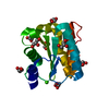
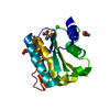
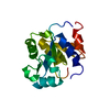
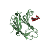
- PDB-3ptj: Structural and functional Analysis of Arabidopsis thaliana thylak... -
+ Open data

- Basic information
| Entry | Database: PDB / ID: 3ptj | ||||||
|---|---|---|---|---|---|---|---|
| Title | Structural and functional Analysis of Arabidopsis thaliana thylakoid lumen protein AtTLP18.3 | ||||||
 Components Components | UPF0603 protein At1g54780, chloroplastic | ||||||
 Keywords Keywords |  HYDROLASE / TAP domain / Rossmann fold / Acid Phosphatase / Arabidopsis thaliana thylakoid lumen HYDROLASE / TAP domain / Rossmann fold / Acid Phosphatase / Arabidopsis thaliana thylakoid lumen | ||||||
| Function / homology |  Function and homology informationphotosystem II repair / thylakoid lumen / chloroplast thylakoid / thylakoid / acid phosphatase activity / chloroplast thylakoid membrane / chloroplast / mRNA binding / nucleus Function and homology informationphotosystem II repair / thylakoid lumen / chloroplast thylakoid / thylakoid / acid phosphatase activity / chloroplast thylakoid membrane / chloroplast / mRNA binding / nucleusSimilarity search - Function | ||||||
| Biological species |  Arabidopsis thaliana (thale cress) Arabidopsis thaliana (thale cress) | ||||||
| Method | X-RAY DIFFRACTION / SYNCHROTRON / SAD / Resolution: 2.6 Å | ||||||
 Authors Authors | Wu, H.Y. / Liu, M.S. / Lin, T.P. / Cheng, Y.S. | ||||||
 Citation Citation | Journal: Plant Physiol. / Year: 2011 Title: Structural and functional assays of AtTLP18.3 identify its novel acid phosphatase activity in thylakoid lumen Authors: Wu, H.Y. / Liu, M.S. / Lin, T.P. / Cheng, Y.S. | ||||||
| History |
|
- Structure visualization
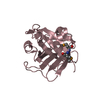
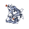
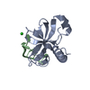
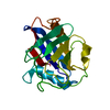
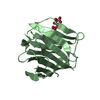
| Structure viewer | Molecule: MolmilJmol/JSmol |
|---|
- Downloads & links
-Download
| PDBx/mmCIF format | 3ptj.cif.gz | 39.1 KB | Display | PDBx/mmCIF format |
|---|---|---|---|---|
| PDB format | pdb3ptj.ent.gz | 30.2 KB | Display | PDB format |
| PDBx/mmJSON format | 3ptj.json.gz | Tree view | PDBx/mmJSON format | |
| Others |  Other downloads Other downloads |
-Validation report
| Arichive directory | https://data.pdbj.org/pub/pdb/validation_reports/pt/3ptjftp://data.pdbj.org/pub/pdb/validation_reports/pt/3ptj | HTTPS FTP |
|---|
-Related structure data


-Links
 PDBj
PDBj- Assembly
| Deposited unit | 
| ||||||||
|---|---|---|---|---|---|---|---|---|---|
| 1 |
| ||||||||
| Unit cell |
|
-Components
| #1: Protein | Mass: 16779.549 Da / Num. of mol.: 1 / Fragment: Phosphatase domain, UNP residues 84-235 / Mutation: L128M, I159M Source method: isolated from a genetically manipulated source Source: (gene. exp.) Arabidopsis thaliana (thale cress) / Strain: cv. Columbia / Gene: At1g54780, T22H22.19 / Plasmid: pGEX6P1 / Production host:  Escherichia coli (E. coli) / Strain (production host): BL21(DE3) / References: UniProt: Q9ZVL6 Escherichia coli (E. coli) / Strain (production host): BL21(DE3) / References: UniProt: Q9ZVL6 |
|---|---|
| #2: Water | ChemComp-HOH / Water Mass: 18.015 Da / Num. of mol.: 67 / Source method: isolated from a natural source / Formula: H2O Mass: 18.015 Da / Num. of mol.: 67 / Source method: isolated from a natural source / Formula: H2O |
-Experimental details
-Experiment
| Experiment | Method: X-RAY DIFFRACTION / Number of used crystals: 1 |
|---|
- Sample preparation
| Crystal | Density Matthews: 2.67 Å3/Da / Density % sol: 54.01 % Description: THE STRUCTURE FACTOR FILE CONTAINS FRIEDEL PAIRS |
|---|---|
| Crystal grow | Temperature: 296 K / Method: vapor diffusion / pH: 6 Details: 0.2M Sodium acetate trihydrate, 0.1M Sodium cacodylate, 25-30% (w/v) Polyethylene glycol 4000, 2mM TCEP , pH 6.0, VAPOR DIFFUSION, temperature 296K |
-Data collection
| Diffraction | Mean temperature: 100 K | |||||||||||||||||||||||||||||||||||||||||||||||||||||||||||||||||||||||||||||
|---|---|---|---|---|---|---|---|---|---|---|---|---|---|---|---|---|---|---|---|---|---|---|---|---|---|---|---|---|---|---|---|---|---|---|---|---|---|---|---|---|---|---|---|---|---|---|---|---|---|---|---|---|---|---|---|---|---|---|---|---|---|---|---|---|---|---|---|---|---|---|---|---|---|---|---|---|---|---|
| Diffraction source | Source: SYNCHROTRON / Site: NSRRC  / Beamline: BL13B1 / Wavelength: 0.97884 Å / Beamline: BL13B1 / Wavelength: 0.97884 Å | |||||||||||||||||||||||||||||||||||||||||||||||||||||||||||||||||||||||||||||
| Detector | Type: ADSC QUANTUM 315r / Detector: CCD / Date: Dec 21, 2008 / Details: MIRRORS | |||||||||||||||||||||||||||||||||||||||||||||||||||||||||||||||||||||||||||||
| Radiation | Monochromator: Si (111) Double crystal / Protocol: SINGLE WAVELENGTH / Monochromatic (M) / Laue (L): M / Scattering type: x-ray | |||||||||||||||||||||||||||||||||||||||||||||||||||||||||||||||||||||||||||||
| Radiation wavelength | Wavelength: 0.97884 Å / Relative weight: 1 | |||||||||||||||||||||||||||||||||||||||||||||||||||||||||||||||||||||||||||||
| Reflection | Resolution: 2.6→30 Å / Num. obs: 11594 / % possible obs: 97.8 % / Observed criterion σ(F): 0 / Observed criterion σ(I): 0 / Redundancy: 9.9 % / Biso Wilson estimate: 33.5 Å2 / Rmerge(I) obs: 0.046 / Rsym value: 0.046 / Net I/σ(I): 30.8 | |||||||||||||||||||||||||||||||||||||||||||||||||||||||||||||||||||||||||||||
| Reflection shell | Diffraction-ID: 1
|
- Processing
| Software |
| |||||||||||||||||||||||||||||||||||||||||||||||||||||||||||||||
|---|---|---|---|---|---|---|---|---|---|---|---|---|---|---|---|---|---|---|---|---|---|---|---|---|---|---|---|---|---|---|---|---|---|---|---|---|---|---|---|---|---|---|---|---|---|---|---|---|---|---|---|---|---|---|---|---|---|---|---|---|---|---|---|---|
| Refinement | Method to determine structure: SAD / Resolution: 2.6→30 Å / Isotropic thermal model: RESTRAINED / Cross valid method: THROUGHOUT / σ(F): 0 / Stereochemistry target values: Engh & Huber / Details: SF file contains Friedel pairs
| |||||||||||||||||||||||||||||||||||||||||||||||||||||||||||||||
| Solvent computation | Bsol: 36.14 Å2 | |||||||||||||||||||||||||||||||||||||||||||||||||||||||||||||||
| Displacement parameters | Biso mean: 32.44 Å2
| |||||||||||||||||||||||||||||||||||||||||||||||||||||||||||||||
| Refine analyze |
| |||||||||||||||||||||||||||||||||||||||||||||||||||||||||||||||
| Refinement step | Cycle: LAST / Resolution: 2.6→30 Å
| |||||||||||||||||||||||||||||||||||||||||||||||||||||||||||||||
| Refine LS restraints |
| |||||||||||||||||||||||||||||||||||||||||||||||||||||||||||||||
| LS refinement shell | Refine-ID: X-RAY DIFFRACTION
| |||||||||||||||||||||||||||||||||||||||||||||||||||||||||||||||
| Xplor file |
|
