 Movie
Movie Controller


+ Open data

- Basic information
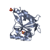
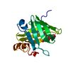
| Entry | Database: PDB / ID: 1t2o | ||||||
|---|---|---|---|---|---|---|---|
| Title | Crystal structure of Se-SrtA, C184-Ala | ||||||
 Components Components | sortase | ||||||
 Keywords Keywords | HYDROLASE / Sortase / BETA BARREL / Transpeptidase | ||||||
| Function / homology |  Function and homology information Function and homology informationcalcium-dependent cysteine-type endopeptidase activity / ion binding / peptide binding / manganese ion binding / calcium ion binding / magnesium ion binding / proteolysisSimilarity search - Function | ||||||
| Biological species |   Staphylococcus aureus (bacteria) Staphylococcus aureus (bacteria) | ||||||
| Method | X-RAY DIFFRACTION / SYNCHROTRON / SAD / Resolution: 2.3 Å | ||||||
 Authors Authors | Zong, Y. / Bice, T.W. / Ton-That, H. / Schneewind, O. / Narayana, S.V. | ||||||
 Citation Citation | Journal: J.Biol.Chem. / Year: 2004 Title: Crystal structures of Staphylococcus aureus sortase A and its substrate complex Authors: Zong, Y. / Bice, T.W. / Ton-That, H. / Schneewind, O. / Narayana, S.V. | ||||||
| History |
|
- Structure visualization
| Structure viewer | Molecule: MolmilJmol/JSmol |
|---|
- Downloads & links
-Download
| PDBx/mmCIF format | 1t2o.cif.gz | 91.3 KB | Display | PDBx/mmCIF format |
|---|---|---|---|---|
| PDB format | pdb1t2o.ent.gz | 74.4 KB | Display | PDB format |
| PDBx/mmJSON format | 1t2o.json.gz | Tree view | PDBx/mmJSON format | |
| Others |  Other downloads Other downloads |
-Validation report
| Arichive directory | https://data.pdbj.org/pub/pdb/validation_reports/t2/1t2oftp://data.pdbj.org/pub/pdb/validation_reports/t2/1t2o | HTTPS FTP |
|---|
-Related structure data


-Links
 PDBj
PDBj- Assembly
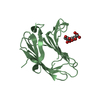
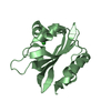
| Deposited unit | 
| ||||||||
|---|---|---|---|---|---|---|---|---|---|
| 1 | 
| ||||||||
| 2 | 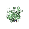
| ||||||||
| 3 | 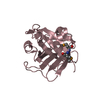
| ||||||||
| Unit cell |
|
-Components
| #1: Protein | Mass: 16555.402 Da / Num. of mol.: 3 / Fragment: Delta-N59 (residues 61-206) / Mutation: C184A Source method: isolated from a genetically manipulated source Source: (gene. exp.) Staphylococcus aureus (bacteria) / Gene: srt / Plasmid: Novagen pET 15b / Production host: Escherichia coli (E. coli) / Strain (production host): XL1-blue / References: UniProt: Q9S446#2: Water | ChemComp-HOH / | Water Mass: 18.015 Da / Num. of mol.: 53 / Source method: isolated from a natural source / Formula: H2O Mass: 18.015 Da / Num. of mol.: 53 / Source method: isolated from a natural source / Formula: H2O |
|---|
-Experimental details
-Experiment
| Experiment | Method: X-RAY DIFFRACTION / Number of used crystals: 1 |
|---|
- Sample preparation
| Crystal | Density Matthews: 1.9 Å3/Da / Density % sol: 40 % / Description: THE FILE CONTAINS FRIEDEL PAIRS |
|---|---|
| Crystal grow | Temperature: 300 K / pH: 6.35 Details: Ammonium Sulfate, MES, Sodium Chloride, pH 6.35, VAPOR DIFFUSION, HANGING DROP, temperature 300K |
-Data collection
| Diffraction | Mean temperature: 100 K | ||||||||||||
|---|---|---|---|---|---|---|---|---|---|---|---|---|---|
| Diffraction source | Source: SYNCHROTRON / Site: NSLS  / Beamline: X4A / Wavelength: 0.9791, 0.9439, 1.212 / Beamline: X4A / Wavelength: 0.9791, 0.9439, 1.212 | ||||||||||||
| Detector | Type: ADSC QUANTUM 4 / Detector: CCD / Date: Mar 20, 2002 | ||||||||||||
| Radiation | Monochromator: NI MIRROR + NI FILTER / Protocol: MAD / Monochromatic (M) / Laue (L): M / Scattering type: x-ray | ||||||||||||
| Radiation wavelength |
| ||||||||||||
| Reflection | Resolution: 2.3→2.4 Å / Num. obs: 35415 / % possible obs: 99 % / Observed criterion σ(I): 2 / Biso Wilson estimate: 23.2 Å2 | ||||||||||||
| Reflection shell | Resolution: 2.3→2.4 Å / % possible all: 99 |
- Processing
| Software |
| ||||||||||||||||||||||||||||||||||||||||||||||||||||||||||||
|---|---|---|---|---|---|---|---|---|---|---|---|---|---|---|---|---|---|---|---|---|---|---|---|---|---|---|---|---|---|---|---|---|---|---|---|---|---|---|---|---|---|---|---|---|---|---|---|---|---|---|---|---|---|---|---|---|---|---|---|---|---|
| Refinement | Method to determine structure: SAD / Resolution: 2.3→17.71 Å / Rfactor Rfree error: 0.005 / Data cutoff high absF: 1604167.94 / Data cutoff low absF: 0 / Isotropic thermal model: GROUP / Cross valid method: THROUGHOUT / σ(F): 0 / Stereochemistry target values: ENGH & HUBER / Details: THE FILE CONTAINS FRIEDEL PAIRS
| ||||||||||||||||||||||||||||||||||||||||||||||||||||||||||||
| Solvent computation | Solvent model: FLAT MODEL / Bsol: 37.53 Å2 / ksol: 0.36 e/Å3 | ||||||||||||||||||||||||||||||||||||||||||||||||||||||||||||
| Displacement parameters | Biso mean: 38.2 Å2
| ||||||||||||||||||||||||||||||||||||||||||||||||||||||||||||
| Refine analyze |
| ||||||||||||||||||||||||||||||||||||||||||||||||||||||||||||
| Refinement step | Cycle: LAST / Resolution: 2.3→17.71 Å
| ||||||||||||||||||||||||||||||||||||||||||||||||||||||||||||
| Refine LS restraints |
| ||||||||||||||||||||||||||||||||||||||||||||||||||||||||||||
| LS refinement shell | Resolution: 2.3→2.44 Å / Rfactor Rfree error: 0.014 / Total num. of bins used: 6
| ||||||||||||||||||||||||||||||||||||||||||||||||||||||||||||
| Xplor file |
|
