 Movie
Movie Controller


+ Open data

- Basic information
| Entry | Database: PDB / ID: 5dlo | ||||||
|---|---|---|---|---|---|---|---|
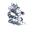
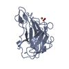
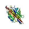
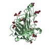
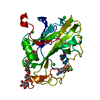
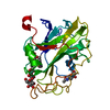
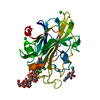
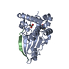
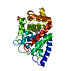
| Title | S. aureus MazF in complex with substrate analogue | ||||||
 Components Components |
| ||||||
 Keywords Keywords |  HYDROLASE / Toxin-Antitoxin / mRNA interferase / ribonuclease / persistence / bacterial stress response HYDROLASE / Toxin-Antitoxin / mRNA interferase / ribonuclease / persistence / bacterial stress response | ||||||
| Function / homology |  Function and homology information Function and homology informationrRNA catabolic process / mRNA catabolic process / RNA endonuclease activity / endonuclease activity / Hydrolases; Acting on ester bonds / DNA binding / RNA bindingSimilarity search - Function | ||||||
| Biological species |   Staphylococcus aureus (bacteria) Staphylococcus aureus (bacteria) Enterobacterio phage MS2 (virus) Enterobacterio phage MS2 (virus) | ||||||
| Method | X-RAY DIFFRACTION / SYNCHROTRON / MOLECULAR REPLACEMENT / Resolution: 1.401 Å | ||||||
 Authors Authors | Zorzini, V. / Loris, R. | ||||||
| Funding support |  Belgium, 1items Belgium, 1items
| ||||||
 Citation Citation | Journal: To Be Published Title: Substrate recognition, regulation mechanism and activity regulation of MazF mRNA interferase. Authors: Zorzini, V. / Loris, R. | ||||||
| History |
|
- Structure visualization
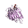
| Structure viewer | Molecule: MolmilJmol/JSmol |
|---|
- Downloads & links
-Download
| PDBx/mmCIF format | 5dlo.cif.gz | 76.3 KB | Display | PDBx/mmCIF format |
|---|---|---|---|---|
| PDB format | pdb5dlo.ent.gz | 54.9 KB | Display | PDB format |
| PDBx/mmJSON format | 5dlo.json.gz | Tree view | PDBx/mmJSON format | |
| Others |  Other downloads Other downloads |
-Validation report
| Arichive directory | https://data.pdbj.org/pub/pdb/validation_reports/dl/5dloftp://data.pdbj.org/pub/pdb/validation_reports/dl/5dlo | HTTPS FTP |
|---|
-Related structure data
| Related structure data |  4mzmS S: Starting model for refinement |
|---|---|
| Similar structure data |
-Links
 PDBj
PDBj- Assembly
| Deposited unit | 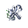
| ||||||||
|---|---|---|---|---|---|---|---|---|---|
| 1 | 
| ||||||||
| Unit cell |
|
-Components
| #1: Protein | Mass: 14952.206 Da / Num. of mol.: 1 Source method: isolated from a genetically manipulated source Source: (gene. exp.) Staphylococcus aureus (bacteria) / Gene: mazF, SAHV_2053 / Production host: Escherichia coli (E. coli)References: UniProt: A7X4P5, UniProt: Q2FWI8*PLUS, Hydrolases; Acting on ester bonds |
|---|---|
| #2: DNA chain | Mass: 2077.385 Da / Num. of mol.: 1 / Source method: obtained synthetically / Source: (synth.) Enterobacterio phage MS2 (virus) |
| #3: Chemical | ChemComp-CL / Chloride  Mass: 35.453 Da / Num. of mol.: 1 / Source method: obtained synthetically / Formula: Cl Mass: 35.453 Da / Num. of mol.: 1 / Source method: obtained synthetically / Formula: Cl |
| #4: Water | ChemComp-HOH / Water Mass: 18.015 Da / Num. of mol.: 195 / Source method: isolated from a natural source / Formula: H2O Mass: 18.015 Da / Num. of mol.: 195 / Source method: isolated from a natural source / Formula: H2O |
-Experimental details
-Experiment
| Experiment | Method: X-RAY DIFFRACTION |
|---|
- Sample preparation
| Crystal | Density Matthews: 2.48 Å3/Da / Density % sol: 50.43 % |
|---|---|
| Crystal grow | Temperature: 292 K / Method: vapor diffusion, hanging drop Details: 75 mM Tris pH 8.5, 18.75% v/v tert-Butanol, 25% Glycerol |
-Data collection
| Diffraction | Mean temperature: 100 K |
|---|---|
| Diffraction source | Source: SYNCHROTRON / Site: SOLEIL  / Beamline: PROXIMA 2 / Wavelength: 0.9801 Å / Beamline: PROXIMA 2 / Wavelength: 0.9801 Å |
| Detector | Type: ADSC QUANTUM 315r / Detector: CCD / Date: Nov 15, 2014 |
| Radiation | Protocol: SINGLE WAVELENGTH / Monochromatic (M) / Laue (L): M / Scattering type: x-ray |
| Radiation wavelength | Wavelength: 0.9801 Å / Relative weight: 1 |
| Reflection | Resolution: 1.4→31.8 Å / Num. obs: 31425 / % possible obs: 99 % / Redundancy: 5.4 % / Rsym value: 0.048 / Net I/σ(I): 21.64 |
| Reflection shell | Resolution: 1.4→1.49 Å / Mean I/σ(I) obs: 2.03 / % possible all: 99 |
- Processing
| Software |
| ||||||||||||||||||||||||||||||||||||||||||||||||||||||||||||||||||||||||||||||||||||
|---|---|---|---|---|---|---|---|---|---|---|---|---|---|---|---|---|---|---|---|---|---|---|---|---|---|---|---|---|---|---|---|---|---|---|---|---|---|---|---|---|---|---|---|---|---|---|---|---|---|---|---|---|---|---|---|---|---|---|---|---|---|---|---|---|---|---|---|---|---|---|---|---|---|---|---|---|---|---|---|---|---|---|---|---|---|
| Refinement | Method to determine structure: MOLECULAR REPLACEMENT Starting model: 4MZM Resolution: 1.401→31.8 Å / SU ML: 0.37 / Cross valid method: THROUGHOUT / σ(F): 1.37 / Phase error: 14.12 / Stereochemistry target values: ML
| ||||||||||||||||||||||||||||||||||||||||||||||||||||||||||||||||||||||||||||||||||||
| Solvent computation | Shrinkage radii: 1.06 Å / VDW probe radii: 1.3 Å / Solvent model: FLAT BULK SOLVENT MODEL / Bsol: 58.992 Å2 / ksol: 0.416 e/Å3 | ||||||||||||||||||||||||||||||||||||||||||||||||||||||||||||||||||||||||||||||||||||
| Displacement parameters |
| ||||||||||||||||||||||||||||||||||||||||||||||||||||||||||||||||||||||||||||||||||||
| Refinement step | Cycle: LAST / Resolution: 1.401→31.8 Å
| ||||||||||||||||||||||||||||||||||||||||||||||||||||||||||||||||||||||||||||||||||||
| Refine LS restraints |
| ||||||||||||||||||||||||||||||||||||||||||||||||||||||||||||||||||||||||||||||||||||
| LS refinement shell |
|
