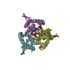
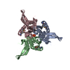
 Movie
Movie Controller

[English] 日本語
 Yorodumi
- PDB-3pcv: Crystal structure analysis of human leukotriene C4 synthase at 1.... -
+ Open data

- Basic information
| Entry | Database: PDB / ID: 3pcv | ||||||
|---|---|---|---|---|---|---|---|
| Title | Crystal structure analysis of human leukotriene C4 synthase at 1.9 angstrom resolution | ||||||
 Components Components | Leukotriene C4 synthase | ||||||
 Keywords Keywords | LYASE / membrane protein / helix bundle / homo trimer / mGST / MAPEG | ||||||
| Function / homology |  Function and homology information Function and homology informationBiosynthesis of protectin and resolvin conjugates in tissue regeneration (PCTR and RCTR) / Biosynthesis of maresin conjugates in tissue regeneration (MCTR) / leukotriene-C4 synthase / leukotriene-C4 synthase activity / Synthesis of Lipoxins (LX) / Synthesis of 5-eicosatetraenoic acids / leukotriene metabolic process / Transferases; Transferring alkyl or aryl groups, other than methyl groups / Synthesis of Leukotrienes (LT) and Eoxins (EX) / leukotriene biosynthetic process ...Biosynthesis of protectin and resolvin conjugates in tissue regeneration (PCTR and RCTR) / Biosynthesis of maresin conjugates in tissue regeneration (MCTR) / leukotriene-C4 synthase / leukotriene-C4 synthase activity / Synthesis of Lipoxins (LX) / Synthesis of 5-eicosatetraenoic acids / leukotriene metabolic process / Transferases; Transferring alkyl or aryl groups, other than methyl groups / Synthesis of Leukotrienes (LT) and Eoxins (EX) / leukotriene biosynthetic process / glutathione peroxidase activity / nuclear outer membrane / long-chain fatty acid biosynthetic process / glutathione transferase activity / enzyme activator activity / nuclear envelope / nuclear membrane / intracellular membrane-bounded organelle / lipid binding / endoplasmic reticulum membrane / endoplasmic reticulum / membrane / identical protein bindingSimilarity search - Function | ||||||
| Biological species |  Homo sapiens (human) Homo sapiens (human) | ||||||
| Method | X-RAY DIFFRACTION / SYNCHROTRON / MOLECULAR REPLACEMENT / Resolution: 1.9 Å | ||||||
 Authors Authors | Saino, H. / Ago, H. / Miyano, M. | ||||||
 Citation Citation | Journal: J.Biol.Chem. / Year: 2011 Title: The catalytic architecture of leukotriene C4 synthase with two arginine residues Authors: Saino, H. / Ukita, Y. / Ago, H. / Irikura, D. / Nisawa, A. / Ueno, G. / Yamamoto, M. / Kanaoka, Y. / Lam, B.K. / Austen, K.F. / Miyano, M. #1: Journal: Nature / Year: 2007Title: Crystal structure of a human membrane protein involved in cysteinyl leukotriene biosynthesis Authors: Ago, H. / Kanaoka, Y. / Irikura, D. / Lam, B.K. / Shimamura, T. / Austen, K.F. / Miyano, M. | ||||||
| History |
|
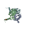
- Structure visualization
| Structure viewer | Molecule: MolmilJmol/JSmol |
|---|
- Downloads & links
-Download
| PDBx/mmCIF format | 3pcv.cif.gz | 54.3 KB | Display | PDBx/mmCIF format |
|---|---|---|---|---|
| PDB format | pdb3pcv.ent.gz | 37.3 KB | Display | PDB format |
| PDBx/mmJSON format | 3pcv.json.gz | Tree view | PDBx/mmJSON format | |
| Others |  Other downloads Other downloads |
-Validation report
| Arichive directory | https://data.pdbj.org/pub/pdb/validation_reports/pc/3pcvftp://data.pdbj.org/pub/pdb/validation_reports/pc/3pcv | HTTPS FTP |
|---|
-Related structure data
| Related structure data |  2uuiS S: Starting model for refinement |
|---|---|
| Similar structure data |

-Links
 PDBj
PDBj


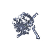
- Assembly
| Deposited unit | 
| ||||||||
|---|---|---|---|---|---|---|---|---|---|
| 1 | 
| ||||||||
| Unit cell |
| ||||||||
| Components on special symmetry positions |
|
-Components
-Protein / Sugars , 2 types, 12 molecules A
| #1: Protein | / LTC4 synthase / Leukotriene-C(4) synthase Mass: 17411.539 Da / Num. of mol.: 1 Source method: isolated from a genetically manipulated source Source: (gene. exp.) Homo sapiens (human) / Gene: LTC4S / Plasmid: pESP / Production host:  Schizosaccharomyces pombe (fission yeast) / References: UniProt: Q16873, leukotriene-C4 synthase Schizosaccharomyces pombe (fission yeast) / References: UniProt: Q16873, leukotriene-C4 synthase |
|---|---|
| #3: Sugar | ChemComp-LMT /  Type: D-saccharide / Mass: 510.615 Da / Num. of mol.: 11 / Source method: obtained synthetically / Formula: C24H46O11 / Comment: detergent*YM Type: D-saccharide / Mass: 510.615 Da / Num. of mol.: 11 / Source method: obtained synthetically / Formula: C24H46O11 / Comment: detergent*YM |
-Non-polymers , 5 types, 105 molecules 


| #2: Chemical | ChemComp-GSH / Glutathione Mass: 307.323 Da / Num. of mol.: 1 / Source method: obtained synthetically / Formula: C10H17N3O6S Mass: 307.323 Da / Num. of mol.: 1 / Source method: obtained synthetically / Formula: C10H17N3O6S | ||||||
|---|---|---|---|---|---|---|---|
| #4: Chemical | ChemComp-EDO / Ethylene glycol Mass: 62.068 Da / Num. of mol.: 4 / Source method: obtained synthetically / Formula: C2H6O2 Mass: 62.068 Da / Num. of mol.: 4 / Source method: obtained synthetically / Formula: C2H6O2#5: Chemical | ChemComp-UNL / Num. of mol.: 4 / Source method: obtained synthetically #6: Chemical | Sulfate Mass: 96.063 Da / Num. of mol.: 2 / Source method: obtained synthetically / Formula: SO4 Mass: 96.063 Da / Num. of mol.: 2 / Source method: obtained synthetically / Formula: SO4#7: Water | ChemComp-HOH / | WaterMass: 18.015 Da / Num. of mol.: 94 / Source method: isolated from a natural source / Formula: H2O |
-Details
| Nonpolymer details | LIGANDS ASSINGED UNL IN THE COORDINATES WERE SUPPOSED TO BE THE MALTOSIDE PORTIONS OF DODECYL ...LIGANDS ASSINGED UNL IN THE COORDINATE |
|---|
-Experimental details
-Experiment
| Experiment | Method: X-RAY DIFFRACTION / Number of used crystals: 2 |
|---|
- Sample preparation
| Crystal | Density Matthews: 4.91 Å3/Da / Density % sol: 74.77 % |
|---|---|
| Crystal grow | Temperature: 293 K / Method: vapor diffusion, sitting drop / pH: 6.5 Details: 0.1M MES-NaOH (pH 6.5), 1.6M ammonium sulfate, 0.8M magnesium chloride, VAPOR DIFFUSION, SITTING DROP, temperature 293K |
-Data collection
| Diffraction | Mean temperature: 100 K |
|---|---|
| Diffraction source | Source: SYNCHROTRON / Site: SPring-8  / Beamline: BL26B2 / Wavelength: 0.97 Å / Beamline: BL26B2 / Wavelength: 0.97 Å |
| Detector | Type: MARMOSAIC 225 mm CCD / Detector: CCD / Date: Sep 5, 2008 |
| Radiation | Monochromator: SAGITALLY FOCUSED Si(111) / Protocol: SINGLE WAVELENGTH / Monochromatic (M) / Laue (L): M / Scattering type: x-ray |
| Radiation wavelength | Wavelength: 0.97 Å / Relative weight: 1 |
| Reflection | Resolution: 1.9→20.4 Å / Num. obs: 30950 / % possible obs: 99.9 % / Observed criterion σ(F): 0 / Observed criterion σ(I): 0 / Redundancy: 9.9 % / Rmerge(I) obs: 0.08 |
| Reflection shell | Resolution: 1.9→2 Å / Redundancy: 7.7 % / Rmerge(I) obs: 0.302 / Num. unique all: 4517 / % possible all: 100 |
- Processing
| Software |
| |||||||||||||||||||||||||||||||||||||||
|---|---|---|---|---|---|---|---|---|---|---|---|---|---|---|---|---|---|---|---|---|---|---|---|---|---|---|---|---|---|---|---|---|---|---|---|---|---|---|---|---|
| Refinement | Method to determine structure: MOLECULAR REPLACEMENT Starting model: PDB ENTRY 2UUI Resolution: 1.9→18.78 Å / Cor.coef. Fo:Fc: 0.958 / Cor.coef. Fo:Fc free: 0.947 / SU B: 1.6 / SU ML: 0.049 / Cross valid method: THROUGHOUT / ESU R Free: 0.084 / Stereochemistry target values: MON_LIB in CCP4
| |||||||||||||||||||||||||||||||||||||||
| Solvent computation | Ion probe radii: 0.8 Å / Shrinkage radii: 0.8 Å / VDW probe radii: 1.4 Å / Solvent model: BABINET MODEL WITH MASK | |||||||||||||||||||||||||||||||||||||||
| Displacement parameters | Biso mean: 29.263 Å2 | |||||||||||||||||||||||||||||||||||||||
| Refinement step | Cycle: LAST / Resolution: 1.9→18.78 Å
| |||||||||||||||||||||||||||||||||||||||
| Refine LS restraints |
| |||||||||||||||||||||||||||||||||||||||
| LS refinement shell | Resolution: 1.901→1.949 Å / Total num. of bins used: 20
|
