 Movie
Movie Controller


+ Open data

- Basic information
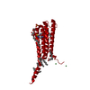
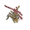
| Entry | Database: PDB / ID: 2uui | ||||||
|---|---|---|---|---|---|---|---|
| Title | Crystal structure of Human Leukotriene C4 Synthase | ||||||
 Components Components | LEUKOTRIENE C4 SYNTHASE | ||||||
 Keywords Keywords | LYASE / LEUKOTRIENE SIGNALLING / LEUKOTRIENE BIOSYNTHESIS / MEMBRANE / EICOSANOID / TRANSMEMBRANE / MEMBRANE PROTEIN / APO / MAPEG / HUMAN / LTC4S / ENZYME / TRIMER | ||||||
| Function / homology |  Function and homology information Function and homology informationBiosynthesis of protectin and resolvin conjugates in tissue regeneration (PCTR and RCTR) / Biosynthesis of maresin conjugates in tissue regeneration (MCTR) / leukotriene-C4 synthase / leukotriene-C4 synthase activity / Synthesis of Lipoxins (LX) / Synthesis of 5-eicosatetraenoic acids / leukotriene metabolic process / Transferases; Transferring alkyl or aryl groups, other than methyl groups / Synthesis of Leukotrienes (LT) and Eoxins (EX) / leukotriene biosynthetic process ...Biosynthesis of protectin and resolvin conjugates in tissue regeneration (PCTR and RCTR) / Biosynthesis of maresin conjugates in tissue regeneration (MCTR) / leukotriene-C4 synthase / leukotriene-C4 synthase activity / Synthesis of Lipoxins (LX) / Synthesis of 5-eicosatetraenoic acids / leukotriene metabolic process / Transferases; Transferring alkyl or aryl groups, other than methyl groups / Synthesis of Leukotrienes (LT) and Eoxins (EX) / leukotriene biosynthetic process / glutathione peroxidase activity / nuclear outer membrane / long-chain fatty acid biosynthetic process / glutathione transferase activity / enzyme activator activity / nuclear envelope / nuclear membrane / intracellular membrane-bounded organelle / lipid binding / endoplasmic reticulum membrane / endoplasmic reticulum / membrane / identical protein bindingSimilarity search - Function | ||||||
| Biological species |  HOMO SAPIENS (human) HOMO SAPIENS (human) | ||||||
| Method | X-RAY DIFFRACTION / SYNCHROTRON / MAD / Resolution: 2 Å | ||||||
 Authors Authors | Martinez Molina, D. / Wetterholm, A. / Kohl, A. / McCarthy, A.A. / Niegowski, D. / Ohlson, E. / Hammarberg, T. / Eshaghi, S. / Haeggstrom, J.Z. / Nordlund, P. | ||||||
 Citation Citation | Journal: Nature / Year: 2007 Title: Structural Basis for Synthesis of Inflammatory Mediators by Human Leukotriene C4 Synthase. Authors: Martinez Molina, D. / Wetterholm, A. / Kohl, A. / Mccarthy, A.A. / Niegowski, D. / Ohlson, E. / Hammarberg, T. / Eshaghi, S. / Haeggstrom, J.Z. / Nordlund, P. | ||||||
| History |
| ||||||
| Remark 650 | HELIX DETERMINATION METHOD: AUTHOR PROVIDED. |
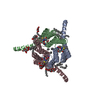
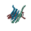
- Structure visualization
| Structure viewer | Molecule: MolmilJmol/JSmol |
|---|
- Downloads & links
-Download
| PDBx/mmCIF format | 2uui.cif.gz | 56.1 KB | Display | PDBx/mmCIF format |
|---|---|---|---|---|
| PDB format | pdb2uui.ent.gz | 41.1 KB | Display | PDB format |
| PDBx/mmJSON format | 2uui.json.gz | Tree view | PDBx/mmJSON format | |
| Others |  Other downloads Other downloads |
-Validation report
| Arichive directory | https://data.pdbj.org/pub/pdb/validation_reports/uu/2uuiftp://data.pdbj.org/pub/pdb/validation_reports/uu/2uui | HTTPS FTP |
|---|
-Related structure data


-Links
 PDBj
PDBj


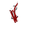
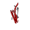
- Assembly
| Deposited unit | 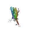
| |||||||||||||||||||||
|---|---|---|---|---|---|---|---|---|---|---|---|---|---|---|---|---|---|---|---|---|---|---|
| 1 | 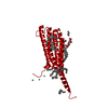
| |||||||||||||||||||||
| Unit cell |
| |||||||||||||||||||||
| Components on special symmetry positions |
|
-Components
-Protein / Sugars , 2 types, 3 molecules A
| #1: Protein | / LEUKOTRIENE-C(4) SYNTHASE / LTC4 SYNTHASE Mass: 17411.545 Da / Num. of mol.: 1 / Fragment: RESIDUES 2-150 Source method: isolated from a genetically manipulated source Source: (gene. exp.) HOMO SAPIENS (human) / Plasmid: PPICZ / Production host:  PICHIA PASTORIS (fungus) / Strain (production host): KM71H / References: UniProt: Q16873, leukotriene-C4 synthase PICHIA PASTORIS (fungus) / Strain (production host): KM71H / References: UniProt: Q16873, leukotriene-C4 synthase |
|---|---|
| #3: Sugar |  Type: D-saccharide / Mass: 510.615 Da / Num. of mol.: 2 / Source method: obtained synthetically / Formula: C24H46O11 / Comment: detergent*YM Type: D-saccharide / Mass: 510.615 Da / Num. of mol.: 2 / Source method: obtained synthetically / Formula: C24H46O11 / Comment: detergent*YM |
-Non-polymers , 4 types, 129 molecules 


| #2: Chemical | Nickel Mass: 58.693 Da / Num. of mol.: 3 / Source method: obtained synthetically / Formula: Ni Mass: 58.693 Da / Num. of mol.: 3 / Source method: obtained synthetically / Formula: Ni#4: Chemical | ChemComp-PLM / Palmitic acid Mass: 256.424 Da / Num. of mol.: 14 / Source method: obtained synthetically / Formula: C16H32O2 Mass: 256.424 Da / Num. of mol.: 14 / Source method: obtained synthetically / Formula: C16H32O2#5: Chemical | Sulfate Mass: 96.063 Da / Num. of mol.: 2 / Source method: obtained synthetically / Formula: SO4 Mass: 96.063 Da / Num. of mol.: 2 / Source method: obtained synthetically / Formula: SO4#6: Water | ChemComp-HOH / | WaterMass: 18.015 Da / Num. of mol.: 110 / Source method: isolated from a natural source / Formula: H2O |
|---|
-Experimental details
-Experiment
| Experiment | Method: X-RAY DIFFRACTION / Number of used crystals: 1 |
|---|
- Sample preparation
| Crystal | Density Matthews: 5.84 Å3/Da / Density % sol: 72.83 % / Description: NONE |
|---|
-Data collection
| Diffraction | Mean temperature: 100 K |
|---|---|
| Diffraction source | Source: SYNCHROTRON / Site: ESRF  / Beamline: ID14-4 / Wavelength: 0.939 / Beamline: ID14-4 / Wavelength: 0.939 |
| Detector | Type: ADSC CCD / Detector: CCD |
| Radiation | Protocol: SINGLE WAVELENGTH / Monochromatic (M) / Laue (L): M / Scattering type: x-ray |
| Radiation wavelength | Wavelength: 0.939 Å / Relative weight: 1 |
| Reflection | Resolution: 1.7→51.16 Å / Num. obs: 44247 / % possible obs: 99.8 % / Redundancy: 7.2 % / Rmerge(I) obs: 0.08 / Net I/σ(I): 16.1 |
| Reflection shell | Resolution: 2→2.11 Å / Redundancy: 7.3 % / Rmerge(I) obs: 0.82 / Mean I/σ(I) obs: 2.2 / % possible all: 100 |
- Processing
| Software | Name: REFMAC / Version: 5.2.0019 / Classification: refinement | ||||||||||||||||||||||||||||||||||||||||||||||||||||||||||||||||||||||||||||||||||||||||||||||||||||||||||||||||||||||||||||||||||||||||||||||||||||||||||||||||||||||||||||||||||||||
|---|---|---|---|---|---|---|---|---|---|---|---|---|---|---|---|---|---|---|---|---|---|---|---|---|---|---|---|---|---|---|---|---|---|---|---|---|---|---|---|---|---|---|---|---|---|---|---|---|---|---|---|---|---|---|---|---|---|---|---|---|---|---|---|---|---|---|---|---|---|---|---|---|---|---|---|---|---|---|---|---|---|---|---|---|---|---|---|---|---|---|---|---|---|---|---|---|---|---|---|---|---|---|---|---|---|---|---|---|---|---|---|---|---|---|---|---|---|---|---|---|---|---|---|---|---|---|---|---|---|---|---|---|---|---|---|---|---|---|---|---|---|---|---|---|---|---|---|---|---|---|---|---|---|---|---|---|---|---|---|---|---|---|---|---|---|---|---|---|---|---|---|---|---|---|---|---|---|---|---|---|---|---|---|
| Refinement | Method to determine structure: MAD Starting model: NONE Resolution: 2→28.27 Å / Cor.coef. Fo:Fc: 0.95 / Cor.coef. Fo:Fc free: 0.936 / SU B: 2.98 / SU ML: 0.084 / Cross valid method: THROUGHOUT / ESU R: 0.114 / ESU R Free: 0.12 / Stereochemistry target values: MAXIMUM LIKELIHOOD / Details: HYDROGENS HAVE BEEN ADDED IN THE RIDING POSITIONS
| ||||||||||||||||||||||||||||||||||||||||||||||||||||||||||||||||||||||||||||||||||||||||||||||||||||||||||||||||||||||||||||||||||||||||||||||||||||||||||||||||||||||||||||||||||||||
| Solvent computation | Ion probe radii: 0.8 Å / Shrinkage radii: 0.8 Å / VDW probe radii: 1.2 Å / Solvent model: MASK | ||||||||||||||||||||||||||||||||||||||||||||||||||||||||||||||||||||||||||||||||||||||||||||||||||||||||||||||||||||||||||||||||||||||||||||||||||||||||||||||||||||||||||||||||||||||
| Displacement parameters | Biso mean: 38.73 Å2 | ||||||||||||||||||||||||||||||||||||||||||||||||||||||||||||||||||||||||||||||||||||||||||||||||||||||||||||||||||||||||||||||||||||||||||||||||||||||||||||||||||||||||||||||||||||||
| Refinement step | Cycle: LAST / Resolution: 2→28.27 Å
| ||||||||||||||||||||||||||||||||||||||||||||||||||||||||||||||||||||||||||||||||||||||||||||||||||||||||||||||||||||||||||||||||||||||||||||||||||||||||||||||||||||||||||||||||||||||
| Refine LS restraints |
|