 Movie
Movie Controller


+ Open data

- Basic information
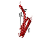
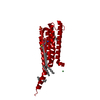
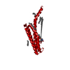
| Entry | Database: PDB / ID: 4ntb | ||||||
|---|---|---|---|---|---|---|---|
| Title | Mus Musculus LTC4 synthase in GSH complex form | ||||||
 Components Components | Leukotriene C4 synthase | ||||||
 Keywords Keywords | LYASE / product analogs / lipid biosynthesis | ||||||
| Function / homology |  Function and homology information Function and homology informationSynthesis of 5-eicosatetraenoic acids / Synthesis of Leukotrienes (LT) and Eoxins (EX) / Synthesis of Lipoxins (LX) / Biosynthesis of maresin conjugates in tissue regeneration (MCTR) / Biosynthesis of protectin and resolvin conjugates in tissue regeneration (PCTR and RCTR) / leukotriene-C4 synthase / leukotriene-C4 synthase activity / leukotriene metabolic process / Transferases; Transferring alkyl or aryl groups, other than methyl groups / glutathione binding ...Synthesis of 5-eicosatetraenoic acids / Synthesis of Leukotrienes (LT) and Eoxins (EX) / Synthesis of Lipoxins (LX) / Biosynthesis of maresin conjugates in tissue regeneration (MCTR) / Biosynthesis of protectin and resolvin conjugates in tissue regeneration (PCTR and RCTR) / leukotriene-C4 synthase / leukotriene-C4 synthase activity / leukotriene metabolic process / Transferases; Transferring alkyl or aryl groups, other than methyl groups / glutathione binding / leukotriene biosynthetic process / glutathione peroxidase activity / nuclear outer membrane / long-chain fatty acid biosynthetic process / glutathione transferase activity / enzyme activator activity / nuclear envelope / nuclear membrane / lipid binding / protein-containing complex binding / endoplasmic reticulum membrane / endoplasmic reticulum / identical protein bindingSimilarity search - Function | ||||||
| Biological species |  Mus musculus (house mouse) Mus musculus (house mouse) | ||||||
| Method | X-RAY DIFFRACTION / SYNCHROTRON / MOLECULAR REPLACEMENT / molecular replacement / Resolution: 2.7 Å | ||||||
 Authors Authors | Niegowski, D. / Rinaldo-Matthis, A. / Haeggstrom, J.Z. | ||||||
 Citation Citation | Journal: To be Published Title: Mus Musculus LTC4 synthase in GSH complex form Authors: Niegowski, D. / Rinaldo-Matthis, A. / Kleinschmidt, T. / Qureshi, A.A. / Haeggstrom, J.Z. | ||||||
| History |
|
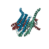
- Structure visualization
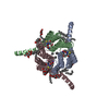
| Structure viewer | Molecule: MolmilJmol/JSmol |
|---|
- Downloads & links
-Download
| PDBx/mmCIF format | 4ntb.cif.gz | 76.8 KB | Display | PDBx/mmCIF format |
|---|---|---|---|---|
| PDB format | pdb4ntb.ent.gz | 56.9 KB | Display | PDB format |
| PDBx/mmJSON format | 4ntb.json.gz | Tree view | PDBx/mmJSON format | |
| Others |  Other downloads Other downloads |
-Validation report
| Arichive directory | https://data.pdbj.org/pub/pdb/validation_reports/nt/4ntbftp://data.pdbj.org/pub/pdb/validation_reports/nt/4ntb | HTTPS FTP |
|---|
-Related structure data
| Related structure data | 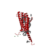 2uuiS S: Starting model for refinement |
|---|---|
| Similar structure data |


-Links
 PDBj
PDBj


- Assembly
| Deposited unit | 
| ||||||||
|---|---|---|---|---|---|---|---|---|---|
| 1 | 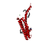
| ||||||||
| Unit cell |
| ||||||||
| Components on special symmetry positions |
|
-Components
| #1: Protein | / LTC4 synthase / Leukotriene-C(4) synthase Mass: 17657.840 Da / Num. of mol.: 1 Source method: isolated from a genetically manipulated source Source: (gene. exp.) Mus musculus (house mouse) / Gene: Ltc4s / Production host:  Komagataella pastoris (fungus) / References: UniProt: Q60860, leukotriene-C4 synthase Komagataella pastoris (fungus) / References: UniProt: Q60860, leukotriene-C4 synthase |
|---|---|
| #2: Chemical | ChemComp-NI / Nickel  Mass: 58.693 Da / Num. of mol.: 1 / Source method: obtained synthetically / Formula: Ni Mass: 58.693 Da / Num. of mol.: 1 / Source method: obtained synthetically / Formula: Ni |
| #3: Chemical | ChemComp-SO4 / Sulfate  Mass: 96.063 Da / Num. of mol.: 1 / Source method: obtained synthetically / Formula: SO4 Mass: 96.063 Da / Num. of mol.: 1 / Source method: obtained synthetically / Formula: SO4 |
| #4: Chemical | ChemComp-GSH / Glutathione  Mass: 307.323 Da / Num. of mol.: 1 / Source method: obtained synthetically / Formula: C10H17N3O6S Mass: 307.323 Da / Num. of mol.: 1 / Source method: obtained synthetically / Formula: C10H17N3O6S |
| #5: Chemical | Palmitic acid  Mass: 256.424 Da / Num. of mol.: 3 / Source method: obtained synthetically / Formula: C16H32O2 Mass: 256.424 Da / Num. of mol.: 3 / Source method: obtained synthetically / Formula: C16H32O2 |
-Experimental details
-Experiment
| Experiment | Method: X-RAY DIFFRACTION / Number of used crystals: 1 |
|---|
- Sample preparation
| Crystal | Density Matthews: 5.72 Å3/Da / Density % sol: 78.51 % |
|---|---|
| Crystal grow | Temperature: 298 K / Method: vapor diffusion / pH: 6.2 Details: 2.1 M NH4SO4, 0.2 M NaCl, 0.1 M Na-cacadylate, pH 6.2, VAPOR DIFFUSION, temperature 298K |
-Data collection
| Diffraction | Mean temperature: 100 K | |||||||||||||||||||||||||||||||||||||||||||||||||||||||||||||||||||||||||||||
|---|---|---|---|---|---|---|---|---|---|---|---|---|---|---|---|---|---|---|---|---|---|---|---|---|---|---|---|---|---|---|---|---|---|---|---|---|---|---|---|---|---|---|---|---|---|---|---|---|---|---|---|---|---|---|---|---|---|---|---|---|---|---|---|---|---|---|---|---|---|---|---|---|---|---|---|---|---|---|
| Diffraction source | Source: SYNCHROTRON / Site: Diamond  / Beamline: I24 / Wavelength: 0.9399 Å / Beamline: I24 / Wavelength: 0.9399 Å | |||||||||||||||||||||||||||||||||||||||||||||||||||||||||||||||||||||||||||||
| Detector | Type: PSI PILATUS 6M / Detector: PIXEL / Date: Jul 6, 2013 | |||||||||||||||||||||||||||||||||||||||||||||||||||||||||||||||||||||||||||||
| Radiation | Protocol: SINGLE WAVELENGTH / Monochromatic (M) / Laue (L): M / Scattering type: x-ray | |||||||||||||||||||||||||||||||||||||||||||||||||||||||||||||||||||||||||||||
| Radiation wavelength | Wavelength: 0.9399 Å / Relative weight: 1 | |||||||||||||||||||||||||||||||||||||||||||||||||||||||||||||||||||||||||||||
| Reflection | Resolution: 1.996→97.959 Å / Num. all: 11166 / Num. obs: 11166 / % possible obs: 99.9 % / Redundancy: 11.1 % / Biso Wilson estimate: 73.41 Å2 / Rsym value: 0.079 / Net I/σ(I): 21.3 | |||||||||||||||||||||||||||||||||||||||||||||||||||||||||||||||||||||||||||||
| Reflection shell |
|
-Phasing
| Phasing | Method: molecular replacement | |||||||||
|---|---|---|---|---|---|---|---|---|---|---|
| Phasing MR |
|
- Processing
| Software |
| ||||||||||||||||||||||||||||||||||||||||||||||||||||||||||||||||||||||||||||||||||||||||||||||||||||
|---|---|---|---|---|---|---|---|---|---|---|---|---|---|---|---|---|---|---|---|---|---|---|---|---|---|---|---|---|---|---|---|---|---|---|---|---|---|---|---|---|---|---|---|---|---|---|---|---|---|---|---|---|---|---|---|---|---|---|---|---|---|---|---|---|---|---|---|---|---|---|---|---|---|---|---|---|---|---|---|---|---|---|---|---|---|---|---|---|---|---|---|---|---|---|---|---|---|---|---|---|---|
| Refinement | Method to determine structure: MOLECULAR REPLACEMENT Starting model: pdb entry 2UUI with water Resolution: 2.7→28.216 Å / Occupancy max: 1 / Occupancy min: 0.33 / SU ML: 0.29 / σ(F): 1.34 / Phase error: 25.01 / Stereochemistry target values: ML
| ||||||||||||||||||||||||||||||||||||||||||||||||||||||||||||||||||||||||||||||||||||||||||||||||||||
| Solvent computation | Shrinkage radii: 0.9 Å / VDW probe radii: 1.11 Å / Solvent model: FLAT BULK SOLVENT MODEL | ||||||||||||||||||||||||||||||||||||||||||||||||||||||||||||||||||||||||||||||||||||||||||||||||||||
| Displacement parameters | Biso mean: 77.7568 Å2 | ||||||||||||||||||||||||||||||||||||||||||||||||||||||||||||||||||||||||||||||||||||||||||||||||||||
| Refinement step | Cycle: LAST / Resolution: 2.7→28.216 Å
| ||||||||||||||||||||||||||||||||||||||||||||||||||||||||||||||||||||||||||||||||||||||||||||||||||||
| Refine LS restraints |
| ||||||||||||||||||||||||||||||||||||||||||||||||||||||||||||||||||||||||||||||||||||||||||||||||||||
| LS refinement shell |
| ||||||||||||||||||||||||||||||||||||||||||||||||||||||||||||||||||||||||||||||||||||||||||||||||||||
| Refinement TLS params. | Method: refined / Refine-ID: X-RAY DIFFRACTION
| ||||||||||||||||||||||||||||||||||||||||||||||||||||||||||||||||||||||||||||||||||||||||||||||||||||
| Refinement TLS group |
|
