 Movie
Movie Controller

[English] 日本語
 Yorodumi
- PDB-6lkw: Structural and functional insights into macrophage migration inhi... -
+ Open data

- Basic information
| Entry | Database: PDB / ID: 6lkw | ||||||
|---|---|---|---|---|---|---|---|
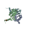
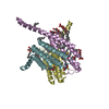
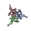
| Title | Structural and functional insights into macrophage migration inhibitory factor from Oncomelania hupensis, the intermediate host of Schistosoma japonicum | ||||||
 Components Components | (Macrophage migration inhibitory ...) x 4 | ||||||
 Keywords Keywords |  CYTOKINE / Oncomelania hupensis macrophage migration inhibitory factor / Isomerase / Beta-alpha-beta fold CYTOKINE / Oncomelania hupensis macrophage migration inhibitory factor / Isomerase / Beta-alpha-beta fold | ||||||
| Function / homology | phenylpyruvate tautomerase / L-dopachrome isomerase / Macrophage migration inhibitory factor / Macrophage migration inhibitory factor (MIF) / Tautomerase/MIF superfamily / cytokine activity / extracellular space / L-dopachrome isomerase Function and homology information Function and homology information | ||||||
| Biological species |  Oncomelania hupensis (invertebrata) Oncomelania hupensis (invertebrata) | ||||||
| Method | X-RAY DIFFRACTION / SYNCHROTRON / MOLECULAR REPLACEMENT / Resolution: 3.2 Å | ||||||
 Authors Authors | Su, Z.M. / Tian, X.Y. / Li, H.J. / Wei, Z.M. / Chen, L.F. / Ren, H.X. / Peng, W.F. / Tang, C.T. | ||||||
 Citation Citation | Journal: Biochem.J. / Year: 2020 Title: Structural and functional insights into macrophage migration inhibitory factor from Oncomelania hupensis, the intermediate host of Schistosoma japonicum. Authors: Su, Z. / Tian, X. / Li, H. / Wei, Z. / Chen, L. / Wang, S. / Ren, H. / Peng, W. / Tang, C. / Lin, T. / Huang, S. | ||||||
| History |
|
- Structure visualization
| Structure viewer | Molecule: MolmilJmol/JSmol |
|---|
- Downloads & links
-Download
| PDBx/mmCIF format | 6lkw.cif.gz | 122.4 KB | Display | PDBx/mmCIF format |
|---|---|---|---|---|
| PDB format | pdb6lkw.ent.gz | 94.3 KB | Display | PDB format |
| PDBx/mmJSON format | 6lkw.json.gz | Tree view | PDBx/mmJSON format | |
| Others |  Other downloads Other downloads |
-Validation report
| Arichive directory | https://data.pdbj.org/pub/pdb/validation_reports/lk/6lkwftp://data.pdbj.org/pub/pdb/validation_reports/lk/6lkw | HTTPS FTP |
|---|
-Related structure data
| Related structure data |  6lkvC  6lr3C 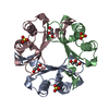 5bsiS S: Starting model for refinement C: citing same article ( |
|---|---|
| Similar structure data |


-Links
 PDBj
PDBj- Assembly
| Deposited unit | 
| ||||||||||||
|---|---|---|---|---|---|---|---|---|---|---|---|---|---|
| 1 | 
| ||||||||||||
| 2 | 
| ||||||||||||
| Unit cell |
| ||||||||||||
| Components on special symmetry positions |
|
-Components
-Macrophage migration inhibitory ... , 4 types, 4 molecules ABDC
| #1: Protein | Mass: 15639.184 Da / Num. of mol.: 1 Source method: isolated from a genetically manipulated source Source: (gene. exp.) Oncomelania hupensis (invertebrata) / Gene: MIF / Production host:  Escherichia coli (E. coli) / References: UniProt: A0A1U9W5E8 Escherichia coli (E. coli) / References: UniProt: A0A1U9W5E8 |
|---|---|
| #2: Protein | Mass: 15710.262 Da / Num. of mol.: 1 Source method: isolated from a genetically manipulated source Source: (gene. exp.) Oncomelania hupensis (invertebrata) / Gene: MIF / Production host: Escherichia coli (E. coli) / References: UniProt: A0A1U9W5E8 |
| #3: Protein | Mass: 15299.792 Da / Num. of mol.: 1 Source method: isolated from a genetically manipulated source Source: (gene. exp.) Oncomelania hupensis (invertebrata) / Gene: MIF / Production host: Escherichia coli (E. coli) / References: UniProt: A0A1U9W5E8 |
| #4: Protein | Mass: 15026.477 Da / Num. of mol.: 1 Source method: isolated from a genetically manipulated source Source: (gene. exp.) Oncomelania hupensis (invertebrata) / Gene: MIF / Production host: Escherichia coli (E. coli) / References: UniProt: A0A1U9W5E8 |
-Non-polymers , 2 types, 116 molecules 

| #5: Chemical | Chloride Mass: 35.453 Da / Num. of mol.: 2 / Source method: obtained synthetically / Formula: Cl / Feature type: SUBJECT OF INVESTIGATION Mass: 35.453 Da / Num. of mol.: 2 / Source method: obtained synthetically / Formula: Cl / Feature type: SUBJECT OF INVESTIGATION#6: Water | ChemComp-HOH / | WaterMass: 18.015 Da / Num. of mol.: 114 / Source method: isolated from a natural source / Formula: H2O |
|---|
-Details
| Has ligand of interest | Y |
|---|---|
| Sequence details | It may be sequencing errors or exist mutations of Oncomelania hupensis. From density map of ...It may be sequencing errors or exist mutations of Oncomelania hupensis. From density map of structure, it seems that residues of number 89, 90, 125 are correct. |
-Experimental details
-Experiment
| Experiment | Method: X-RAY DIFFRACTION / Number of used crystals: 1 |
|---|
- Sample preparation
| Crystal | Density Matthews: 3.49 Å3/Da / Density % sol: 64.76 % |
|---|---|
| Crystal grow | Temperature: 289 K / Method: vapor diffusion, sitting drop / pH: 4.6 Details: 30% (w/v) MPD, 0.1 M Sodium acetate (pH 4.6), and 20 mM Calcium chloride |
-Data collection
| Diffraction | Mean temperature: 100 K / Serial crystal experiment: N |
|---|---|
| Diffraction source | Source: SYNCHROTRON / Site: SSRF  / Beamline: BL19U1 / Wavelength: 0.97855 Å / Beamline: BL19U1 / Wavelength: 0.97855 Å |
| Detector | Type: PSI PILATUS 6M / Detector: PIXEL / Date: Jun 30, 2019 |
| Radiation | Protocol: SINGLE WAVELENGTH / Monochromatic (M) / Laue (L): M / Scattering type: x-ray |
| Radiation wavelength | Wavelength: 0.97855 Å / Relative weight: 1 |
| Reflection | Resolution: 3.2→50 Å / Num. obs: 15176 / % possible obs: 100 % / Redundancy: 75.7 % / CC star: 0.998 / Rmerge(I) obs: 0.1 / Net I/σ(I): 55.33 |
| Reflection shell | Resolution: 3.2→3.26 Å / Redundancy: 65.2 % / Rmerge(I) obs: 0.596 / Mean I/σ(I) obs: 10 / Num. unique obs: 727 / CC star: 0.997 / % possible all: 100 |
- Processing
| Software |
| ||||||||||||||||||||||||||||||||||||||||||||||||||||||||||||
|---|---|---|---|---|---|---|---|---|---|---|---|---|---|---|---|---|---|---|---|---|---|---|---|---|---|---|---|---|---|---|---|---|---|---|---|---|---|---|---|---|---|---|---|---|---|---|---|---|---|---|---|---|---|---|---|---|---|---|---|---|---|
| Refinement | Method to determine structure: MOLECULAR REPLACEMENT Starting model: 5BSI Resolution: 3.2→49.95 Å / Cor.coef. Fo:Fc: 0.909 / Cor.coef. Fo:Fc free: 0.8 / SU B: 28.773 / SU ML: 0.477 / Cross valid method: THROUGHOUT / σ(F): 0 / ESU R Free: 0.6 Details: HYDROGENS HAVE BEEN ADDED IN THE RIDING POSITIONS U VALUES : REFINED INDIVIDUALLY
| ||||||||||||||||||||||||||||||||||||||||||||||||||||||||||||
| Solvent computation | Ion probe radii: 0.8 Å / Shrinkage radii: 0.8 Å / VDW probe radii: 1.2 Å | ||||||||||||||||||||||||||||||||||||||||||||||||||||||||||||
| Displacement parameters | Biso max: 160.99 Å2 / Biso mean: 60.119 Å2 / Biso min: 7.91 Å2
| ||||||||||||||||||||||||||||||||||||||||||||||||||||||||||||
| Refinement step | Cycle: final / Resolution: 3.2→49.95 Å
| ||||||||||||||||||||||||||||||||||||||||||||||||||||||||||||
| Refine LS restraints |
| ||||||||||||||||||||||||||||||||||||||||||||||||||||||||||||
| LS refinement shell | Resolution: 3.2→3.282 Å / Rfactor Rfree error: 0
|
