Resolution: 2→59.03 Å / Num. obs: 19001 / % possible obs: 98.9 % / Redundancy: 12.1 % / Rmerge(I) obs: 0.076 / Χ2: 1.065 / Net I/σ(I): 7.2
Reflection shell
Resolution (Å)
Redundancy (%)
Rmerge(I) obs
Num. unique all
Χ2
Diffraction-ID
% possible all
2-2.07
11.1
0.526
1862
0.393
1
100
2.07-2.15
13.3
0.37
1881
0.412
1
100
2.15-2.25
13.4
0.282
1881
0.528
1
100
2.25-2.37
13.5
0.204
1893
0.617
1
99.9
2.37-2.52
13.8
0.162
1892
0.795
1
100
2.52-2.71
13.4
0.12
1885
0.966
1
99.9
2.71-2.99
13
0.095
1912
1.242
1
99.8
2.99-3.42
11.7
0.077
1913
1.929
1
99.2
3.42-4.31
9.6
0.059
1916
2.403
1
97.3
4.31-50
8.2
0.045
1966
2.2
1
93.7
-
Processing
Software
Name
Version
Classification
NB
DENZO
datareduction
SCALEPACK
datascaling
REFMAC
5.5.0109
refinement
PDB_EXTRACT
3.1
dataextraction
PHENIX
1.7.1_743
refinement
Refinement
Resolution: 2→25.794 Å / SU B: 10.538 / SU ML: 0.68 / Cross valid method: THROUGHOUT / σ(F): 0 / Phase error: 22.15 / Stereochemistry target values: ML Details: HYDROGENS HAVE BEEN ADDED IN THE RIDING POSITIONS U VALUES
Rfactor
Num. reflection
% reflection
Selection details
Rfree
0.2449
964
5.09 %
RANDOM
Rwork
0.1918
-
-
-
obs
0.1942
17987
98.86 %
-
Solvent computation
Shrinkage radii: 0.95 Å / VDW probe radii: 1.2 Å / Solvent model: FLAT BULK SOLVENT MODEL / Bsol: 39.039 Å2 / ksol: 0.324 e/Å3
Displacement parameters
Baniso -1
Baniso -2
Baniso -3
1-
-1.0254 Å2
-0 Å2
0 Å2
2-
-
-1.0254 Å2
-0 Å2
3-
-
-
2.0508 Å2
Refinement step
Cycle: LAST / Resolution: 2→25.794 Å
Protein
Nucleic acid
Ligand
Solvent
Total
Num. atoms
1969
0
20
101
2090
Refine LS restraints
Refine-ID
Type
Dev ideal
Number
X-RAY DIFFRACTION
f_bond_d
0.003
2047
X-RAY DIFFRACTION
f_angle_d
0.643
2777
X-RAY DIFFRACTION
f_dihedral_angle_d
14.188
733
X-RAY DIFFRACTION
f_chiral_restr
0.045
292
X-RAY DIFFRACTION
f_plane_restr
0.003
366
LS refinement shell
Refine-ID: X-RAY DIFFRACTION / Total num. of bins used: 7
Resolution (Å)
Rfactor Rfree
Num. reflection Rfree
Rfactor Rwork
Num. reflection Rwork
% reflection obs (%)
1.9999-2.1053
0.3535
134
0.3027
2531
99
2.1053-2.2372
0.3235
154
0.2446
2527
100
2.2372-2.4098
0.3074
138
0.2285
2537
100
2.4098-2.6521
0.2657
163
0.2229
2545
100
2.6521-3.0353
0.268
138
0.2082
2597
100
3.0353-3.8222
0.2504
137
0.1841
2582
99
3.8222-25.796
0.1798
100
0.1637
2665
95
Refinement TLS params.
Method: refined / Origin x: -9.6529 Å / Origin y: -20.4148 Å / Origin z: -21.1565 Å
11
12
13
21
22
23
31
32
33
T
0.2387 Å2
0.0355 Å2
0.0781 Å2
-
0.3681 Å2
0.0911 Å2
-
-
0.3292 Å2
L
2.6626 °2
0.413 °2
0.2045 °2
-
3.9208 °2
-1.2449 °2
-
-
4.5134 °2
S
0.2017 Å °
0.0022 Å °
0.1711 Å °
-0.2827 Å °
0.0408 Å °
0.1056 Å °
0.0718 Å °
-0.308 Å °
-0.1825 Å °
Refinement TLS group
Selection details: all
+
About Yorodumi
-
News
-
Feb 9, 2022. New format data for meta-information of EMDB entries
New format data for meta-information of EMDB entries
Version 3 of the EMDB header file is now the official format.
The previous official version 1.9 will be removed from the archive.
In the structure databanks used in Yorodumi, some data are registered as the other names, "COVID-19 virus" and "2019-nCoV". Here are the details of the virus and the list of structure data.
Jan 31, 2019. EMDB accession codes are about to change! (news from PDBe EMDB page)
EMDB accession codes are about to change! (news from PDBe EMDB page)
The allocation of 4 digits for EMDB accession codes will soon come to an end. Whilst these codes will remain in use, new EMDB accession codes will include an additional digit and will expand incrementally as the available range of codes is exhausted. The current 4-digit format prefixed with “EMD-” (i.e. EMD-XXXX) will advance to a 5-digit format (i.e. EMD-XXXXX), and so on. It is currently estimated that the 4-digit codes will be depleted around Spring 2019, at which point the 5-digit format will come into force.
The EM Navigator/Yorodumi systems omit the EMD- prefix.
Related info.:Q: What is EMD? / ID/Accession-code notation in Yorodumi/EM Navigator
Yorodumi is a browser for structure data from EMDB, PDB, SASBDB, etc.
This page is also the successor to EM Navigator detail page, and also detail information page/front-end page for Omokage search.
The word "yorodu" (or yorozu) is an old Japanese word meaning "ten thousand". "mi" (miru) is to see.
Related info.:EMDB / PDB / SASBDB / Comparison of 3 databanks / Yorodumi Search / Aug 31, 2016. New EM Navigator & Yorodumi / Yorodumi Papers / Jmol/JSmol / Function and homology information / Changes in new EM Navigator and Yorodumi
 Movie
Movie Controller


 Open data
 Basic information
 Components
 Keywords
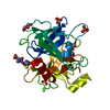
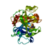
 Function and homology information
 matriptase / epithelial cell morphogenesis involved in placental branching /

 Authors
 Citation
 Structure visualization
 Downloads & links
 Other downloads


 PDBj
PDBj


 Assembly
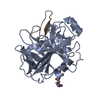

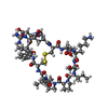
 Type: Polypeptide
 Mass: 307.323 Da / Num. of mol.: 1 / Source method: obtained synthetically / Formula: C10H17N3O6S
 Mass: 18.015 Da / Num. of mol.: 101 / Source method: isolated from a natural source / Formula: H2O
 Sample preparation
 / Beamline: 22-ID / Wavelength: 1.04 Å
 Processing