 Movie
Movie Controller

[English] 日本語
 Yorodumi
- PDB-4irq: Crystal structure of catalytic domain of human beta1,4galactosylt... -
+ Open data

- Basic information
| Entry | Database: PDB / ID: 4irq | ||||||
|---|---|---|---|---|---|---|---|
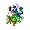
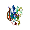
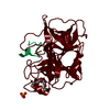
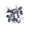
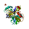
| Title | Crystal structure of catalytic domain of human beta1,4galactosyltransferase 7 in closed conformation in complex with manganese and UDP | ||||||
 Components Components | Beta-1,4-galactosyltransferase 7 | ||||||
 Keywords Keywords |  TRANSFERASE / GT-A fold / closed conformation / manganese and UDP complex / glycosyltransferase / Golgi TRANSFERASE / GT-A fold / closed conformation / manganese and UDP complex / glycosyltransferase / Golgi | ||||||
| Function / homology |  Function and homology informationxylosylprotein 4-beta-galactosyltransferase / xylosylprotein 4-beta-galactosyltransferase activity / glycosylation / galactosyltransferase activity / glycosaminoglycan metabolic process / Defective B4GALT7 causes EDS, progeroid type / beta-N-acetylglucosaminylglycopeptide beta-1,4-galactosyltransferase activity / A tetrasaccharide linker sequence is required for GAG synthesis / glycosaminoglycan biosynthetic process / proteoglycan biosynthetic process ...xylosylprotein 4-beta-galactosyltransferase / xylosylprotein 4-beta-galactosyltransferase activity / glycosylation / galactosyltransferase activity / glycosaminoglycan metabolic process / Defective B4GALT7 causes EDS, progeroid type / beta-N-acetylglucosaminylglycopeptide beta-1,4-galactosyltransferase activity / A tetrasaccharide linker sequence is required for GAG synthesis / glycosaminoglycan biosynthetic process / proteoglycan biosynthetic process / proteoglycan metabolic process / protein N-linked glycosylation / Transferases; Glycosyltransferases; Hexosyltransferases / Golgi cisterna membrane / supramolecular fiber organization / negative regulation of fibroblast proliferation / protein modification process / manganese ion binding / carbohydrate metabolic process / Golgi membrane / Golgi apparatus / membrane Function and homology informationxylosylprotein 4-beta-galactosyltransferase / xylosylprotein 4-beta-galactosyltransferase activity / glycosylation / galactosyltransferase activity / glycosaminoglycan metabolic process / Defective B4GALT7 causes EDS, progeroid type / beta-N-acetylglucosaminylglycopeptide beta-1,4-galactosyltransferase activity / A tetrasaccharide linker sequence is required for GAG synthesis / glycosaminoglycan biosynthetic process / proteoglycan biosynthetic process ...xylosylprotein 4-beta-galactosyltransferase / xylosylprotein 4-beta-galactosyltransferase activity / glycosylation / galactosyltransferase activity / glycosaminoglycan metabolic process / Defective B4GALT7 causes EDS, progeroid type / beta-N-acetylglucosaminylglycopeptide beta-1,4-galactosyltransferase activity / A tetrasaccharide linker sequence is required for GAG synthesis / glycosaminoglycan biosynthetic process / proteoglycan biosynthetic process / proteoglycan metabolic process / protein N-linked glycosylation / Transferases; Glycosyltransferases; Hexosyltransferases / Golgi cisterna membrane / supramolecular fiber organization / negative regulation of fibroblast proliferation / protein modification process / manganese ion binding / carbohydrate metabolic process / Golgi membrane / Golgi apparatus / membraneSimilarity search - Function | ||||||
| Biological species |  Homo sapiens (human) Homo sapiens (human) | ||||||
| Method | X-RAY DIFFRACTION / MOLECULAR REPLACEMENT / Resolution: 2.3 Å | ||||||
 Authors Authors | Tsutsui, Y. / Ramakrishnan, B. / Qasba, P.K. | ||||||
 Citation Citation | Journal: J.Biol.Chem. / Year: 2013 Title: Crystal structures of beta-1,4-galactosyltransferase 7 enzyme reveal conformational changes and substrate binding. Authors: Tsutsui, Y. / Ramakrishnan, B. / Qasba, P.K. | ||||||
| History |
|
- Structure visualization
| Structure viewer | Molecule: MolmilJmol/JSmol |
|---|
- Downloads & links
-Download
| PDBx/mmCIF format | 4irq.cif.gz | 221.4 KB | Display | PDBx/mmCIF format |
|---|---|---|---|---|
| PDB format | pdb4irq.ent.gz | 176.5 KB | Display | PDB format |
| PDBx/mmJSON format | 4irq.json.gz | Tree view | PDBx/mmJSON format | |
| Others |  Other downloads Other downloads |
-Validation report
| Arichive directory | https://data.pdbj.org/pub/pdb/validation_reports/ir/4irqftp://data.pdbj.org/pub/pdb/validation_reports/ir/4irq | HTTPS FTP |
|---|
-Related structure data
| Related structure data |  4irpC 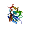 4lw3C  4lw6C  4m4kC  3lw6S C: citing same article ( S: Starting model for refinement |
|---|---|
| Similar structure data |


-Links
 PDBj
PDBj


- Assembly
| Deposited unit | 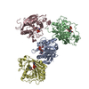
| ||||||||
|---|---|---|---|---|---|---|---|---|---|
| 1 | 
| ||||||||
| 2 | 
| ||||||||
| 3 | 
| ||||||||
| 4 | 
| ||||||||
| 5 | 
| ||||||||
| 6 | 
| ||||||||
| Unit cell |
|
-Components
| #1: Protein | Mass: 29216.273 Da / Num. of mol.: 4 Source method: isolated from a genetically manipulated source Details: human Galectin-1 as fusion protein / Source: (gene. exp.) Homo sapiens (human)Gene: 4galactosyltransferase, B4GALT7, beta1, UNQ748/PRO1478, XGALT1 Plasmid: PET23a / Production host:  Escherichia coli (E. coli) / Strain (production host): BL21 Escherichia coli (E. coli) / Strain (production host): BL21References: UniProt: Q9UBV7, Transferases; Glycosyltransferases; Hexosyltransferases, xylosylprotein 4-beta-galactosyltransferase#2: Chemical | ChemComp-MN /   Mass: 54.938 Da / Num. of mol.: 4 / Source method: obtained synthetically / Formula: Mn Mass: 54.938 Da / Num. of mol.: 4 / Source method: obtained synthetically / Formula: Mn#3: Chemical | ChemComp-UDP / Uridine diphosphate  Type: RNA linking / Mass: 404.161 Da / Num. of mol.: 4 / Source method: obtained synthetically / Formula: C9H14N2O12P2 / Comment: UDP*YM Type: RNA linking / Mass: 404.161 Da / Num. of mol.: 4 / Source method: obtained synthetically / Formula: C9H14N2O12P2 / Comment: UDP*YM#4: Chemical | ChemComp-TRS / Tris  Mass: 122.143 Da / Num. of mol.: 4 / Source method: obtained synthetically / Formula: C4H12NO3 / Comment: pH buffer*YM Mass: 122.143 Da / Num. of mol.: 4 / Source method: obtained synthetically / Formula: C4H12NO3 / Comment: pH buffer*YM#5: Water | ChemComp-HOH / | Water Mass: 18.015 Da / Num. of mol.: 289 / Source method: isolated from a natural source / Formula: H2O Mass: 18.015 Da / Num. of mol.: 289 / Source method: isolated from a natural source / Formula: H2O |
|---|
-Experimental details
-Experiment
| Experiment | Method: X-RAY DIFFRACTION / Number of used crystals: 1 |
|---|
- Sample preparation
| Crystal | Density Matthews: 3.38 Å3/Da / Density % sol: 63.57 % |
|---|---|
| Crystal grow | Temperature: 291 K / Method: vapor diffusion, hanging drop / pH: 8.5 Details: 100mM Tri.HCl, pH 8.5, 8% PEG 8000 as precipitating agent, VAPOR DIFFUSION, HANGING DROP, temperature 291K |
-Data collection
| Diffraction | Mean temperature: 100 K |
|---|---|
| Diffraction source | Source: ROTATING ANODE / Type: RIGAKU MICROMAX-007 HF / Wavelength: 1.5418 Å |
| Detector | Type: MAR scanner 345 mm plate / Detector: IMAGE PLATE / Date: Aug 27, 2012 / Details: mirrors |
| Radiation | Monochromator: Graphite / Protocol: SINGLE WAVELENGTH / Monochromatic (M) / Laue (L): M / Scattering type: x-ray |
| Radiation wavelength | Wavelength: 1.5418 Å / Relative weight: 1 |
| Reflection | Resolution: 2.3→50 Å / Num. all: 66815 / % possible obs: 94 % / Observed criterion σ(I): 1 / Redundancy: 4.4 % / Rsym value: 0.091 / Net I/σ(I): 13.8 |
| Reflection shell | Resolution: 2.3→2.38 Å / Redundancy: 2.7 % / Mean I/σ(I) obs: 2 / Num. unique all: 5725 / Rsym value: 0.523 / % possible all: 83.3 |
- Processing
| Software |
| |||||||||||||||||||||||||||||||||||||||||||||||||||||||||||||||||||||||||||||||||||||||||||||||||||||||||||||||||||||||||||||||||||||||||||||||||||||||||||||||||||||||||||||||
|---|---|---|---|---|---|---|---|---|---|---|---|---|---|---|---|---|---|---|---|---|---|---|---|---|---|---|---|---|---|---|---|---|---|---|---|---|---|---|---|---|---|---|---|---|---|---|---|---|---|---|---|---|---|---|---|---|---|---|---|---|---|---|---|---|---|---|---|---|---|---|---|---|---|---|---|---|---|---|---|---|---|---|---|---|---|---|---|---|---|---|---|---|---|---|---|---|---|---|---|---|---|---|---|---|---|---|---|---|---|---|---|---|---|---|---|---|---|---|---|---|---|---|---|---|---|---|---|---|---|---|---|---|---|---|---|---|---|---|---|---|---|---|---|---|---|---|---|---|---|---|---|---|---|---|---|---|---|---|---|---|---|---|---|---|---|---|---|---|---|---|---|---|---|---|---|---|
| Refinement | Method to determine structure: MOLECULAR REPLACEMENT Starting model: 3LW6 Resolution: 2.3→37.847 Å / SU ML: 0.27 / Cross valid method: THROUGHOUT / σ(F): 0 / Phase error: 27.95 / Stereochemistry target values: ML
| |||||||||||||||||||||||||||||||||||||||||||||||||||||||||||||||||||||||||||||||||||||||||||||||||||||||||||||||||||||||||||||||||||||||||||||||||||||||||||||||||||||||||||||||
| Solvent computation | Shrinkage radii: 0.9 Å / VDW probe radii: 1.11 Å / Solvent model: FLAT BULK SOLVENT MODEL | |||||||||||||||||||||||||||||||||||||||||||||||||||||||||||||||||||||||||||||||||||||||||||||||||||||||||||||||||||||||||||||||||||||||||||||||||||||||||||||||||||||||||||||||
| Refinement step | Cycle: LAST / Resolution: 2.3→37.847 Å
| |||||||||||||||||||||||||||||||||||||||||||||||||||||||||||||||||||||||||||||||||||||||||||||||||||||||||||||||||||||||||||||||||||||||||||||||||||||||||||||||||||||||||||||||
| Refine LS restraints |
| |||||||||||||||||||||||||||||||||||||||||||||||||||||||||||||||||||||||||||||||||||||||||||||||||||||||||||||||||||||||||||||||||||||||||||||||||||||||||||||||||||||||||||||||
| LS refinement shell |
|
