 Movie
Movie Controller


+ Open data

- Basic information
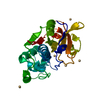
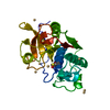
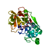
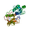
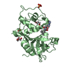
| Entry | Database: PDB / ID: 3p5u | ||||||
|---|---|---|---|---|---|---|---|
| Title | Actinidin from Actinidia arguta planch (Sarusashi) | ||||||
 Components Components | Actinidin Actinidain Actinidain | ||||||
 Keywords Keywords | HYDROLASE / SAD / Cysteine proteinases | ||||||
| Function / homology |  Function and homology information Function and homology information | ||||||
| Biological species |  Actinidia arguta (plant) Actinidia arguta (plant) | ||||||
| Method | X-RAY DIFFRACTION / SAD / Resolution: 1.5 Å | ||||||
 Authors Authors | Manickam, Y. / Nirmal, N. / Suzuki, A. / Sugiyama, Y. / Yamane, T. / Devadasan, V. / Sharma, A. | ||||||
 Citation Citation | Journal: Acta Crystallogr.,Sect.D / Year: 2010 Title: Structural analysis of actinidin and a comparison of cadmium and sulfur anomalous signals from actinidin crystals measured using in-house copper- and chromium-anode X-ray sources Authors: Yogavel, M. / Nithya, N. / Suzuki, A. / Sugiyama, Y. / Yamane, T. / Velmurugan, D. / Sharma, A. | ||||||
| History |
|
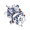
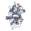
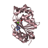
- Structure visualization
| Structure viewer | Molecule: MolmilJmol/JSmol |
|---|
- Downloads & links
-Download
| PDBx/mmCIF format | 3p5u.cif.gz | 59 KB | Display | PDBx/mmCIF format |
|---|---|---|---|---|
| PDB format | pdb3p5u.ent.gz | 46.5 KB | Display | PDB format |
| PDBx/mmJSON format | 3p5u.json.gz | Tree view | PDBx/mmJSON format | |
| Others |  Other downloads Other downloads |
-Validation report
| Arichive directory | https://data.pdbj.org/pub/pdb/validation_reports/p5/3p5uftp://data.pdbj.org/pub/pdb/validation_reports/p5/3p5u | HTTPS FTP |
|---|
-Related structure data


-Links
 PDBj
PDBj
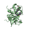
- Assembly
| Deposited unit | 
| ||||||||
|---|---|---|---|---|---|---|---|---|---|
| 1 |
| ||||||||
| Unit cell |
|
-Components
| #1: Protein | Actinidain Mass: 24009.309 Da / Num. of mol.: 1 / Source method: isolated from a natural source / Source: (natural) Actinidia arguta (plant) / References: UniProt: A5HII2*PLUS, actinidain | ||||
|---|---|---|---|---|---|
| #2: Chemical | ChemComp-CD /   Mass: 112.411 Da / Num. of mol.: 5 / Source method: obtained synthetically / Formula: Cd Mass: 112.411 Da / Num. of mol.: 5 / Source method: obtained synthetically / Formula: Cd#3: Water | ChemComp-HOH / | Water Mass: 18.015 Da / Num. of mol.: 288 / Source method: isolated from a natural source / Formula: H2O Mass: 18.015 Da / Num. of mol.: 288 / Source method: isolated from a natural source / Formula: H2OSequence details | A SEQUENCE DATABASE REFERENCE FOR THIS PROTEIN DOES NOT CURRENTLY EXIST. THIS SEQUENCE WILL BE ...A SEQUENCE DATABASE REFERENCE FOR THIS PROTEIN DOES NOT CURRENTLY EXIST. THIS SEQUENCE WILL BE DEPOSITED IN THE SEQUENCE DATABASE. | |
-Experimental details
-Experiment
| Experiment | Method: X-RAY DIFFRACTION / Number of used crystals: 1 |
|---|
- Sample preparation
| Crystal | Density Matthews: 2.02 Å3/Da / Density % sol: 39.19 % |
|---|---|
| Crystal grow | Temperature: 288 K / Method: vapor diffusion, hanging drop / pH: 6 Details: 30% PEG 400, 0.1M sodium acetate, 30mM Cadmium chloride, pH 6.0, VAPOR DIFFUSION, HANGING DROP, temperature 288K |
-Data collection
| Diffraction | Mean temperature: 100 K |
|---|---|
| Diffraction source | Source: ROTATING ANODE / Type: RIGAKU FR-E DW / Wavelength: 1.5418 Å |
| Detector | Type: RIGAKU RAXIS IV / Detector: IMAGE PLATE / Date: Sep 15, 2008 |
| Radiation | Monochromator: Mirrors / Protocol: SINGLE WAVELENGTH / Monochromatic (M) / Laue (L): M / Scattering type: x-ray |
| Radiation wavelength | Wavelength: 1.5418 Å / Relative weight: 1 |
| Reflection | Resolution: 1.5→110 Å / Num. obs: 31698 / % possible obs: 99.5 % / Observed criterion σ(F): 2 / Observed criterion σ(I): 2 / Redundancy: 7 % / Rmerge(I) obs: 0.038 / Net I/σ(I): 45.4 |
| Reflection shell | Resolution: 1.5→1.55 Å / Rmerge(I) obs: 0.251 / Mean I/σ(I) obs: 9.64 / % possible all: 98.4 |
-Phasing
| Phasing | Method: SAD |
|---|
- Processing
| Software |
| ||||||||||||||||||||||||||||||||||||||||||||||||||||||||||||||||||||||||||||||||||||||||||
|---|---|---|---|---|---|---|---|---|---|---|---|---|---|---|---|---|---|---|---|---|---|---|---|---|---|---|---|---|---|---|---|---|---|---|---|---|---|---|---|---|---|---|---|---|---|---|---|---|---|---|---|---|---|---|---|---|---|---|---|---|---|---|---|---|---|---|---|---|---|---|---|---|---|---|---|---|---|---|---|---|---|---|---|---|---|---|---|---|---|---|---|
| Refinement | Method to determine structure: SAD / Resolution: 1.5→18.42 Å / Cor.coef. Fo:Fc: 0.963 / Cor.coef. Fo:Fc free: 0.951 / WRfactor Rfree: 0.2255 / WRfactor Rwork: 0.2021 / Occupancy max: 1 / Occupancy min: 0.25 / FOM work R set: 0.8711 / SU B: 1.289 / SU ML: 0.05 / SU R Cruickshank DPI: 0.0864 / SU Rfree: 0.0831 / Cross valid method: THROUGHOUT / σ(F): 0 / ESU R Free: 0.083 / Stereochemistry target values: MAXIMUM LIKELIHOOD
| ||||||||||||||||||||||||||||||||||||||||||||||||||||||||||||||||||||||||||||||||||||||||||
| Solvent computation | Ion probe radii: 0.8 Å / Shrinkage radii: 0.8 Å / VDW probe radii: 1.2 Å / Solvent model: MASK | ||||||||||||||||||||||||||||||||||||||||||||||||||||||||||||||||||||||||||||||||||||||||||
| Displacement parameters | Biso max: 79.08 Å2 / Biso mean: 17.1737 Å2 / Biso min: 8.72 Å2
| ||||||||||||||||||||||||||||||||||||||||||||||||||||||||||||||||||||||||||||||||||||||||||
| Refinement step | Cycle: LAST / Resolution: 1.5→18.42 Å
| ||||||||||||||||||||||||||||||||||||||||||||||||||||||||||||||||||||||||||||||||||||||||||
| Refine LS restraints |
| ||||||||||||||||||||||||||||||||||||||||||||||||||||||||||||||||||||||||||||||||||||||||||
| LS refinement shell | Resolution: 1.5→1.539 Å / Total num. of bins used: 20
|
