 Movie
Movie Controller


+ Open data

- Basic information
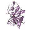
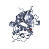
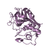
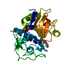
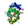
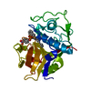
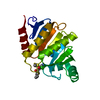
| Entry | Database: PDB / ID: 1aec | ||||||
|---|---|---|---|---|---|---|---|
| Title | CRYSTAL STRUCTURE OF ACTINIDIN-E-64 COMPLEX+ | ||||||
 Components Components | ACTINIDIN Actinidain Actinidain | ||||||
 Keywords Keywords | HYDROLASE | ||||||
| Function / homology |  Function and homology informationactinidain / proteolysis involved in protein catabolic process / lysosome / cysteine-type endopeptidase activity / extracellular space Function and homology informationactinidain / proteolysis involved in protein catabolic process / lysosome / cysteine-type endopeptidase activity / extracellular spaceSimilarity search - Function | ||||||
| Biological species |  Actinidia chinensis (golden kiwifruit) Actinidia chinensis (golden kiwifruit) | ||||||
| Method | X-RAY DIFFRACTION / Resolution: 1.86 Å | ||||||
 Authors Authors | Varughese, K.I. | ||||||
 Citation Citation | Journal: Biochemistry / Year: 1992 Title: Crystal structure of an actinidin-E-64 complex. Authors: Varughese, K.I. / Su, Y. / Cromwell, D. / Hasnain, S. / Xuong, N.H. | ||||||
| History |
|
- Structure visualization
| Structure viewer | Molecule: MolmilJmol/JSmol |
|---|
- Downloads & links
-Download
| PDBx/mmCIF format | 1aec.cif.gz | 67.2 KB | Display | PDBx/mmCIF format |
|---|---|---|---|---|
| PDB format | pdb1aec.ent.gz | 48.9 KB | Display | PDB format |
| PDBx/mmJSON format | 1aec.json.gz | Tree view | PDBx/mmJSON format | |
| Others |  Other downloads Other downloads |
-Validation report
| Arichive directory | https://data.pdbj.org/pub/pdb/validation_reports/ae/1aecftp://data.pdbj.org/pub/pdb/validation_reports/ae/1aec | HTTPS FTP |
|---|
-Related structure data
| Similar structure data |
|---|


-Links
 PDBj
PDBj
- Assembly
| Deposited unit | 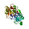
| ||||||||
|---|---|---|---|---|---|---|---|---|---|
| 1 |
| ||||||||
| Unit cell |
| ||||||||
| Atom site foot note | 1: CIS PROLINE - PRO 155 2: THE SIDE CHAIN OF SER 9 IS MODELLED WITH AN ALTERNATE CONFORMATION DUE TO DISORDER. |
-Components
| #1: Protein | Actinidain Mass: 23569.971 Da / Num. of mol.: 1 Source method: isolated from a genetically manipulated source Source: (gene. exp.) Actinidia chinensis (golden kiwifruit) / References: UniProt: P00785, actinidain |
|---|---|
| #2: Chemical | ChemComp-E64 /   Mass: 360.429 Da / Num. of mol.: 1 / Source method: obtained synthetically / Formula: C15H30N5O5 Mass: 360.429 Da / Num. of mol.: 1 / Source method: obtained synthetically / Formula: C15H30N5O5 |
| #3: Water | ChemComp-HOH / Water Mass: 18.015 Da / Num. of mol.: 268 / Source method: isolated from a natural source / Formula: H2O Mass: 18.015 Da / Num. of mol.: 268 / Source method: isolated from a natural source / Formula: H2O |
| Sequence details | SEQUENCE ADVISORY NOTICE: DIFFERENCE BETWEEN PIR AND PDB SEQUENCE. PIR ENTRY NAME: PIR:S12618 ...SEQUENCE ADVISORY NOTICE: DIFFERENCE |
-Experimental details
-Experiment
| Experiment | Method: X-RAY DIFFRACTION |
|---|
- Sample preparation
| Crystal | Density Matthews: 2.24 Å3/Da / Density % sol: 45.13 % | ||||||||||||||||||||||||||||||
|---|---|---|---|---|---|---|---|---|---|---|---|---|---|---|---|---|---|---|---|---|---|---|---|---|---|---|---|---|---|---|---|
| Crystal grow | *PLUS Temperature: 4 ℃ / Method: vapor diffusion, hanging drop | ||||||||||||||||||||||||||||||
| Components of the solutions | *PLUS
|
-Data collection
| Radiation | Scattering type: x-ray |
|---|---|
| Radiation wavelength | Relative weight: 1 |
| Reflection | *PLUS Highest resolution: 1.86 Å / Num. obs: 96911 / % possible obs: 94 % / Num. measured all: 17408 |
- Processing
| Software |
| ||||||||||||||||||||||||||||||||||||||||||||||||||||||||||||
|---|---|---|---|---|---|---|---|---|---|---|---|---|---|---|---|---|---|---|---|---|---|---|---|---|---|---|---|---|---|---|---|---|---|---|---|---|---|---|---|---|---|---|---|---|---|---|---|---|---|---|---|---|---|---|---|---|---|---|---|---|---|
| Refinement | Resolution: 1.86→8 Å / Rfactor Rwork: 0.145 / Rfactor obs: 0.145 / σ(I): 2 | ||||||||||||||||||||||||||||||||||||||||||||||||||||||||||||
| Refinement step | Cycle: LAST / Resolution: 1.86→8 Å
| ||||||||||||||||||||||||||||||||||||||||||||||||||||||||||||
| Refine LS restraints |
| ||||||||||||||||||||||||||||||||||||||||||||||||||||||||||||
| Refinement | *PLUS Highest resolution: 1.86 Å / Lowest resolution: 8 Å / Num. reflection obs: 17116 / σ(I): 2 / Rfactor obs: 0.145 | ||||||||||||||||||||||||||||||||||||||||||||||||||||||||||||
| Solvent computation | *PLUS | ||||||||||||||||||||||||||||||||||||||||||||||||||||||||||||
| Displacement parameters | *PLUS | ||||||||||||||||||||||||||||||||||||||||||||||||||||||||||||
| Refine LS restraints | *PLUS Type: x_angle_d |
