 Movie
Movie Controller

[English] 日本語
 Yorodumi
- PDB-3p3d: Crystal structure of the Nup53 RRM domain from Pichia guilliermondii -
+ Open data

- Basic information
| Entry | Database: PDB / ID: 3p3d | ||||||
|---|---|---|---|---|---|---|---|
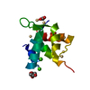
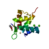
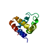
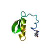
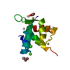
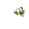
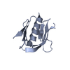
| Title | Crystal structure of the Nup53 RRM domain from Pichia guilliermondii | ||||||
 Components Components | Nucleoporin 53 | ||||||
 Keywords Keywords | NUCLEAR PROTEIN / STRUCTURAL GENOMICS / PSI-2 / PROTEIN STRUCTURE INITIATIVE / NEW YORK STRUCTURAL GENOMIX RESEARCH CONSORTIUM / NYSGXRC / Protein Transport / component of Nuclear Pore Complex / New York SGX Research Center for Structural Genomics | ||||||
| Function / homology |  Function and homology information Function and homology informationstructural constituent of nuclear pore / mRNA transport / nuclear pore / protein transport / nuclear membrane / nucleic acid bindingSimilarity search - Function | ||||||
| Biological species |  Pichia guilliermondii (fungus) Pichia guilliermondii (fungus) | ||||||
| Method | X-RAY DIFFRACTION / SYNCHROTRON / MOLECULAR REPLACEMENT / Resolution: 2.35 Å | ||||||
 Authors Authors | Sampathkumar, P. / Shawn, C. / Bain, K. / Gilmore, J. / Gheyi, T. / Atwell, S. / Thompson, D.A. / Emtage, J.S. / Wasserman, S. / Sauder, J.M. ...Sampathkumar, P. / Shawn, C. / Bain, K. / Gilmore, J. / Gheyi, T. / Atwell, S. / Thompson, D.A. / Emtage, J.S. / Wasserman, S. / Sauder, J.M. / Burley, S.K. / New York SGX Research Center for Structural Genomics (NYSGXRC) | ||||||
 Citation Citation | Journal: To be Published Title: Crystal structure of the Nup53 RRM domain from Pichia guilliermondii Authors: Sampathkumar, P. | ||||||
| History |
|
- Structure visualization
| Structure viewer | Molecule: MolmilJmol/JSmol |
|---|
- Downloads & links
-Download
| PDBx/mmCIF format | 3p3d.cif.gz | 31.1 KB | Display | PDBx/mmCIF format |
|---|---|---|---|---|
| PDB format | pdb3p3d.ent.gz | 19.1 KB | Display | PDB format |
| PDBx/mmJSON format | 3p3d.json.gz | Tree view | PDBx/mmJSON format | |
| Others |  Other downloads Other downloads |
-Validation report
| Arichive directory | https://data.pdbj.org/pub/pdb/validation_reports/p3/3p3dftp://data.pdbj.org/pub/pdb/validation_reports/p3/3p3d | HTTPS FTP |
|---|
-Related structure data
| Related structure data | 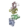 1wwhS S: Starting model for refinement |
|---|---|
| Similar structure data | |
| Other databases |
-Links
 PDBj
PDBj- Assembly
| Deposited unit | 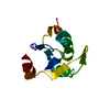
| ||||||||
|---|---|---|---|---|---|---|---|---|---|
| 1 |
| ||||||||
| 2 | 
| ||||||||
| Unit cell |
|
-Components
| #1: Protein | Mass: 14517.317 Da / Num. of mol.: 1 / Fragment: RRM Domain residues 261-383 Source method: isolated from a genetically manipulated source Source: (gene. exp.) Pichia guilliermondii (fungus) / Gene: NUP53 (gi146415668), PGUG_04532 / Plasmid: BC-pSGX3(BC); modified pET26b / Production host:  Escherichia coli (E. coli) / Strain (production host): BL21(DE3)-Codon+RIL / References: UniProt: A5DMN1 Escherichia coli (E. coli) / Strain (production host): BL21(DE3)-Codon+RIL / References: UniProt: A5DMN1 |
|---|---|
| #2: Water | ChemComp-HOH / Water Mass: 18.015 Da / Num. of mol.: 20 / Source method: isolated from a natural source / Formula: H2O Mass: 18.015 Da / Num. of mol.: 20 / Source method: isolated from a natural source / Formula: H2O |
-Experimental details
-Experiment
| Experiment | Method: X-RAY DIFFRACTION / Number of used crystals: 1 |
|---|
- Sample preparation
| Crystal | Density Matthews: 2.14 Å3/Da / Density % sol: 42.55 % |
|---|---|
| Crystal grow | Temperature: 294 K / Method: vapor diffusion, sitting drop / pH: 5.4 Details: 100mM sodium acetate, 20% PEG 1500, pH 5.4, VAPOR DIFFUSION, SITTING DROP, temperature 294K |
-Data collection
| Diffraction | Mean temperature: 100 K |
|---|---|
| Diffraction source | Source: SYNCHROTRON / Site: APS  / Beamline: 31-ID / Wavelength: 0.97929 Å / Beamline: 31-ID / Wavelength: 0.97929 Å |
| Detector | Type: RAYONIX MX225HE / Detector: CCD / Date: Jun 24, 2010 |
| Radiation | Monochromator: diamond / Protocol: SINGLE WAVELENGTH / Monochromatic (M) / Laue (L): M / Scattering type: x-ray |
| Radiation wavelength | Wavelength: 0.97929 Å / Relative weight: 1 |
| Reflection | Resolution: 2.35→38.62 Å / Num. obs: 5437 / % possible obs: 99.8 % / Redundancy: 21 % / Biso Wilson estimate: 59 Å2 / Rsym value: 0.085 / Net I/σ(I): 21.1 |
| Reflection shell | Resolution: 2.35→2.48 Å / Redundancy: 21.9 % / Mean I/σ(I) obs: 6.5 / Num. unique all: 782 / Rsym value: 0.406 / % possible all: 100 |
- Processing
| Software |
| |||||||||||||||||||||||||||||||||||||||||||||||||||||||||||||||||||||||||||||||||||||
|---|---|---|---|---|---|---|---|---|---|---|---|---|---|---|---|---|---|---|---|---|---|---|---|---|---|---|---|---|---|---|---|---|---|---|---|---|---|---|---|---|---|---|---|---|---|---|---|---|---|---|---|---|---|---|---|---|---|---|---|---|---|---|---|---|---|---|---|---|---|---|---|---|---|---|---|---|---|---|---|---|---|---|---|---|---|---|
| Refinement | Method to determine structure: MOLECULAR REPLACEMENT Starting model: Poly-Ala of 1WWH Resolution: 2.35→20.46 Å / Cor.coef. Fo:Fc: 0.935 / Cor.coef. Fo:Fc free: 0.91 / Occupancy max: 1 / Occupancy min: 1 / SU B: 8.577 / SU ML: 0.202 / Cross valid method: THROUGHOUT / σ(F): 0 / ESU R: 0.353 / ESU R Free: 0.273 / Stereochemistry target values: MAXIMUM LIKELIHOOD Details: HYDROGENS HAVE BEEN ADDED IN THE RIDING POSITIONS U VALUES : REFINED INDIVIDUALLY
| |||||||||||||||||||||||||||||||||||||||||||||||||||||||||||||||||||||||||||||||||||||
| Solvent computation | Ion probe radii: 0.8 Å / Shrinkage radii: 0.8 Å / VDW probe radii: 1.4 Å / Solvent model: BABINET MODEL WITH MASK | |||||||||||||||||||||||||||||||||||||||||||||||||||||||||||||||||||||||||||||||||||||
| Displacement parameters | Biso max: 75.06 Å2 / Biso mean: 45.3917 Å2 / Biso min: 29.77 Å2
| |||||||||||||||||||||||||||||||||||||||||||||||||||||||||||||||||||||||||||||||||||||
| Refinement step | Cycle: LAST / Resolution: 2.35→20.46 Å
| |||||||||||||||||||||||||||||||||||||||||||||||||||||||||||||||||||||||||||||||||||||
| Refine LS restraints |
| |||||||||||||||||||||||||||||||||||||||||||||||||||||||||||||||||||||||||||||||||||||
| LS refinement shell | Resolution: 2.353→2.413 Å / Total num. of bins used: 20
|
