 Movie
Movie Controller


+ Open data

- Basic information
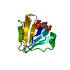
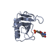
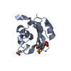
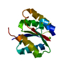
| Entry | Database: PDB / ID: 2v76 | ||||||
|---|---|---|---|---|---|---|---|
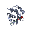
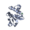
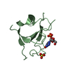
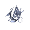
| Title | Crystal structure of the human dok1 PTB domain | ||||||
 Components Components | DOCKING PROTEIN 1 | ||||||
 Keywords Keywords |  PROTEIN BINDING / PROTEIN-BINDING / PTB DOMAIN / PHOSPHORYLATION / ADAPTOR PROTEIN PROTEIN BINDING / PROTEIN-BINDING / PTB DOMAIN / PHOSPHORYLATION / ADAPTOR PROTEIN | ||||||
| Function / homology |  Function and homology information Function and homology informationmacrophage colony-stimulating factor signaling pathway / positive regulation of epidermal growth factor receptor signaling pathway / RET signaling / PTK6 Regulates RTKs and Their Effectors AKT1 and DOK1 / cell surface receptor protein tyrosine kinase signaling pathway / axon guidance / Ras protein signal transduction / cell surface receptor signaling pathway / perinuclear region of cytoplasm / signal transduction ...macrophage colony-stimulating factor signaling pathway / positive regulation of epidermal growth factor receptor signaling pathway / RET signaling / PTK6 Regulates RTKs and Their Effectors AKT1 and DOK1 / cell surface receptor protein tyrosine kinase signaling pathway / axon guidance / Ras protein signal transduction / cell surface receptor signaling pathway / perinuclear region of cytoplasm / signal transduction / nucleus / cytosolSimilarity search - Function | ||||||
| Biological species |  HOMO SAPIENS (human) HOMO SAPIENS (human) | ||||||
| Method | X-RAY DIFFRACTION / SYNCHROTRON / MOLECULAR REPLACEMENT / Resolution: 1.6 Å | ||||||
 Authors Authors | Oxley, C.L. / Anthis, N.J. / Lowe, E.D. / Campbell, I.D. / Wegener, K.L. | ||||||
 Citation Citation | Journal: J.Biol.Chem. / Year: 2008 Title: An Integrin Phosphorylation Switch: The Effect of {Beta}3 Integrin Tail Phosphorylation on Dok1 and Talin Binding. Authors: Oxley, C.L. / Anthis, N.J. / Lowe, E.D. / Vakonakis, I. / Campbell, I.D. / Wegener, K.L. | ||||||
| History |
|
- Structure visualization
| Structure viewer | Molecule: MolmilJmol/JSmol |
|---|
- Downloads & links
-Download
| PDBx/mmCIF format | 2v76.cif.gz | 187.6 KB | Display | PDBx/mmCIF format |
|---|---|---|---|---|
| PDB format | pdb2v76.ent.gz | 152.2 KB | Display | PDB format |
| PDBx/mmJSON format | 2v76.json.gz | Tree view | PDBx/mmJSON format | |
| Others |  Other downloads Other downloads |
-Validation report
| Arichive directory | https://data.pdbj.org/pub/pdb/validation_reports/v7/2v76ftp://data.pdbj.org/pub/pdb/validation_reports/v7/2v76 | HTTPS FTP |
|---|
-Related structure data
| Related structure data |  1p5tS S: Starting model for refinement |
|---|---|
| Similar structure data |


-Links
 PDBj
PDBj


- Assembly
| Deposited unit | 
| ||||||||
|---|---|---|---|---|---|---|---|---|---|
| 1 | 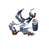
| ||||||||
| 2 | 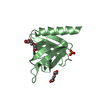
| ||||||||
| 3 | 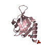
| ||||||||
| 4 | 
| ||||||||
| Unit cell |
|
-Components
-Protein , 1 types, 4 molecules ABCD
| #1: Protein | Mass: 12014.623 Da / Num. of mol.: 4 / Fragment: PTB DOMAIN, RESIDUES 152-256 Source method: isolated from a genetically manipulated source Source: (gene. exp.) HOMO SAPIENS (human) / Production host:  ESCHERICHIA COLI (E. coli) / References: UniProt: Q53TY2, UniProt: Q99704*PLUS ESCHERICHIA COLI (E. coli) / References: UniProt: Q53TY2, UniProt: Q99704*PLUS |
|---|
-Non-polymers , 5 types, 397 molecules 


| #2: Chemical | ChemComp-PGE / Polyethylene glycol Mass: 150.173 Da / Num. of mol.: 4 / Source method: obtained synthetically / Formula: C6H14O4 Mass: 150.173 Da / Num. of mol.: 4 / Source method: obtained synthetically / Formula: C6H14O4#3: Chemical | ChemComp-SO4 / Sulfate Mass: 96.063 Da / Num. of mol.: 4 / Source method: obtained synthetically / Formula: SO4 Mass: 96.063 Da / Num. of mol.: 4 / Source method: obtained synthetically / Formula: SO4#4: Chemical | ChemComp-EDO / | Ethylene glycol Mass: 62.068 Da / Num. of mol.: 1 / Source method: obtained synthetically / Formula: C2H6O2 Mass: 62.068 Da / Num. of mol.: 1 / Source method: obtained synthetically / Formula: C2H6O2#5: Chemical | ChemComp-GOL / | Glycerol Mass: 92.094 Da / Num. of mol.: 1 / Source method: obtained synthetically / Formula: C3H8O3 Mass: 92.094 Da / Num. of mol.: 1 / Source method: obtained synthetically / Formula: C3H8O3#6: Water | ChemComp-HOH / | WaterMass: 18.015 Da / Num. of mol.: 387 / Source method: isolated from a natural source / Formula: H2O |
|---|
-Experimental details
-Experiment
| Experiment | Method: X-RAY DIFFRACTION / Number of used crystals: 1 |
|---|
- Sample preparation
| Crystal | Density Matthews: 2.39 Å3/Da / Density % sol: 48.6 % / Description: NONE |
|---|---|
| Crystal grow | pH: 7.5 Details: 2M AMONIUM SULFATE, 2% PEG 400, 0.1M NA HEPES PH 7.5 |
-Data collection
| Diffraction | Mean temperature: 100 K |
|---|---|
| Diffraction source | Source: SYNCHROTRON / Site: ESRF  / Beamline: ID14-2 / Wavelength: 0.933 / Beamline: ID14-2 / Wavelength: 0.933 |
| Detector | Date: Sep 2, 2006 |
| Radiation | Protocol: SINGLE WAVELENGTH / Monochromatic (M) / Laue (L): M / Scattering type: x-ray |
| Radiation wavelength | Wavelength: 0.933 Å / Relative weight: 1 |
| Reflection | Resolution: 1.6→25.98 Å / Num. obs: 59364 / % possible obs: 99.7 % / Observed criterion σ(I): 2 / Redundancy: 7.5 % / Rmerge(I) obs: 0.06 / Net I/σ(I): 22.5 |
| Reflection shell | Resolution: 1.6→1.68 Å / Redundancy: 3.6 % / Rmerge(I) obs: 0.38 / Mean I/σ(I) obs: 2.5 / % possible all: 98.3 |
- Processing
| Software |
| ||||||||||||||||||||||||||||||||||||||||||||||||||||||||||||||||||||||||||||||||||||||||||||||||||||||||||||||||||||||||||||||||||||||||||||||||||||||||||||||||||||||||||||||||||||||
|---|---|---|---|---|---|---|---|---|---|---|---|---|---|---|---|---|---|---|---|---|---|---|---|---|---|---|---|---|---|---|---|---|---|---|---|---|---|---|---|---|---|---|---|---|---|---|---|---|---|---|---|---|---|---|---|---|---|---|---|---|---|---|---|---|---|---|---|---|---|---|---|---|---|---|---|---|---|---|---|---|---|---|---|---|---|---|---|---|---|---|---|---|---|---|---|---|---|---|---|---|---|---|---|---|---|---|---|---|---|---|---|---|---|---|---|---|---|---|---|---|---|---|---|---|---|---|---|---|---|---|---|---|---|---|---|---|---|---|---|---|---|---|---|---|---|---|---|---|---|---|---|---|---|---|---|---|---|---|---|---|---|---|---|---|---|---|---|---|---|---|---|---|---|---|---|---|---|---|---|---|---|---|---|
| Refinement | Method to determine structure: MOLECULAR REPLACEMENT Starting model: PDB ENTRY 1P5T Resolution: 1.6→59.66 Å / Cor.coef. Fo:Fc: 0.957 / Cor.coef. Fo:Fc free: 0.941 / SU B: 3.871 / SU ML: 0.063 / Cross valid method: THROUGHOUT / ESU R: 0.123 / ESU R Free: 0.094 / Stereochemistry target values: MAXIMUM LIKELIHOOD / Details: HYDROGENS HAVE BEEN ADDED IN THE RIDING POSITIONS.
| ||||||||||||||||||||||||||||||||||||||||||||||||||||||||||||||||||||||||||||||||||||||||||||||||||||||||||||||||||||||||||||||||||||||||||||||||||||||||||||||||||||||||||||||||||||||
| Solvent computation | Ion probe radii: 0.8 Å / Shrinkage radii: 0.8 Å / VDW probe radii: 1.4 Å / Solvent model: MASK | ||||||||||||||||||||||||||||||||||||||||||||||||||||||||||||||||||||||||||||||||||||||||||||||||||||||||||||||||||||||||||||||||||||||||||||||||||||||||||||||||||||||||||||||||||||||
| Displacement parameters | Biso mean: 21.57 Å2
| ||||||||||||||||||||||||||||||||||||||||||||||||||||||||||||||||||||||||||||||||||||||||||||||||||||||||||||||||||||||||||||||||||||||||||||||||||||||||||||||||||||||||||||||||||||||
| Refinement step | Cycle: LAST / Resolution: 1.6→59.66 Å
| ||||||||||||||||||||||||||||||||||||||||||||||||||||||||||||||||||||||||||||||||||||||||||||||||||||||||||||||||||||||||||||||||||||||||||||||||||||||||||||||||||||||||||||||||||||||
| Refine LS restraints |
|