 Movie
Movie Controller

[English] 日本語
 Yorodumi
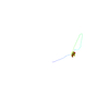
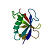
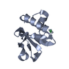
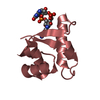
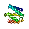
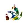
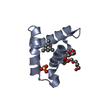
- PDB-1muz: NMR STRUCTURE OF THE TUMOR SUPPRESSOR BIN1: ALTERNATIVE SPLICING ... -
+ Open data

- Basic information
| Entry | Database: PDB / ID: 1muz | ||||||
|---|---|---|---|---|---|---|---|
| Title | NMR STRUCTURE OF THE TUMOR SUPPRESSOR BIN1: ALTERNATIVE SPLICING IN MELANOMA AND INTERACTION WITH C-MYC | ||||||
 Components Components | Myc box dependent interacting protein 1 | ||||||
 Keywords Keywords | ENDOCYTOSIS/EXOCYTOSIS /  TUMOR SUPPRESSOR / ENDOCYTOSIS-EXOCYTOSIS COMPLEX TUMOR SUPPRESSOR / ENDOCYTOSIS-EXOCYTOSIS COMPLEX | ||||||
| Function / homology |  Function and homology information Function and homology informationlipid tube / negative regulation of ventricular cardiac muscle cell action potential / negative regulation of calcium ion transmembrane transport via high voltage-gated calcium channel / negative regulation of aspartic-type endopeptidase activity involved in amyloid precursor protein catabolic process / lipid tube assembly / RNA polymerase II transcription repressor complex / regulation of cell cycle process / T-tubule organization / negative regulation of potassium ion transmembrane transport / varicosity ...lipid tube / negative regulation of ventricular cardiac muscle cell action potential / negative regulation of calcium ion transmembrane transport via high voltage-gated calcium channel / negative regulation of aspartic-type endopeptidase activity involved in amyloid precursor protein catabolic process / lipid tube assembly / RNA polymerase II transcription repressor complex / regulation of cell cycle process / T-tubule organization / negative regulation of potassium ion transmembrane transport / varicosity / extrinsic component of synaptic vesicle membrane / axon initial segment / cerebellar mossy fiber / positive regulation of astrocyte differentiation / nucleus localization / node of Ranvier / aspartic-type endopeptidase inhibitor activity / I band / RNA polymerase binding / nucleus organization / regulation of neuron differentiation / clathrin binding / positive regulation of actin filament polymerization / endosome to lysosome transport / regulation of heart rate by cardiac conduction / positive regulation of endocytosis / negative regulation of amyloid-beta formation / synaptic vesicle endocytosis / axon terminus / cytoskeleton organization / T-tubule / phospholipid binding / tau protein binding / Z disc / endocytosis / actin filament binding / synaptic vesicle / actin cytoskeleton / GTPase binding / Clathrin-mediated endocytosis / nuclear envelope / protein-folding chaperone binding / protease binding / vesicle / endosome / positive regulation of apoptotic process / axon / dendrite / glutamatergic synapse / negative regulation of transcription by RNA polymerase II / membrane / identical protein binding / nucleus / plasma membrane / cytosol / cytoplasmSimilarity search - Function | ||||||
| Biological species |  Homo sapiens (human) Homo sapiens (human) | ||||||
| Method | SOLUTION NMR / torsion angle dynamics | ||||||
 Authors Authors | Pineda-Lucena, A. / Arrowsmith, C.H. | ||||||
 Citation Citation | Journal: J.Mol.Biol. / Year: 2005 Title: A structure-based model of the c-Myc/Bin1 protein interaction shows alternative splicing of Bin1 and c-Myc phosphorylation are key binding determinants. Authors: Pineda-Lucena, A. / Ho, C.S. / Mao, D.Y. / Sheng, Y. / Laister, R.C. / Muhandiram, R. / Lu, Y. / Seet, B.T. / Katz, S. / Szyperski, T. / Penn, L.Z. / Arrowsmith, C.H. | ||||||
| History |
| ||||||
| Remark 999 | SEQUENCE The protein crystallized by the author contains Lys465 which corresponds to Glu576 in the ...SEQUENCE The protein crystallized by the author contains Lys465 which corresponds to Glu576 in the Swiss-Prot entry O00499. This residue conflict is noted in the database reference. |
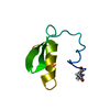
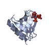
- Structure visualization
| Structure viewer | Molecule: MolmilJmol/JSmol |
|---|
- Downloads & links
-Download
| PDBx/mmCIF format | 1muz.cif.gz | 499.7 KB | Display | PDBx/mmCIF format |
|---|---|---|---|---|
| PDB format | pdb1muz.ent.gz | 434.7 KB | Display | PDB format |
| PDBx/mmJSON format | 1muz.json.gz | Tree view | PDBx/mmJSON format | |
| Others |  Other downloads Other downloads |
-Validation report
| Arichive directory | https://data.pdbj.org/pub/pdb/validation_reports/mu/1muzftp://data.pdbj.org/pub/pdb/validation_reports/mu/1muz | HTTPS FTP |
|---|
-Related structure data


-Links
 PDBj
PDBj


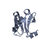
- Assembly
| Deposited unit | 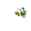
| |||||||||
|---|---|---|---|---|---|---|---|---|---|---|
| 1 |
| |||||||||
| NMR ensembles |
|
-Components
| #1: Protein | Mass: 9356.479 Da / Num. of mol.: 1 / Fragment: residues 513-593 Source method: isolated from a genetically manipulated source Source: (gene. exp.) Homo sapiens (human) / Plasmid: pET15b / Production host:  Escherichia coli (E. coli) / References: UniProt: O00499 Escherichia coli (E. coli) / References: UniProt: O00499 |
|---|
-Experimental details
-Experiment
| Experiment | Method: SOLUTION NMR | ||||||||||||
|---|---|---|---|---|---|---|---|---|---|---|---|---|---|
| NMR experiment |
| ||||||||||||
| NMR details | Text: THIS STRUCTURE WAS DETERMINED USING STANDARD 3D HETERONUCLEAR NMR TECHNIQUES |
- Sample preparation
| Details | Contents: 1.5 mM Bin1(402-482) U-15N, 13C 25 mM sodium phosphate buffer, 150 mM NaCl, 1 mM DTT, 95% H2O, 5% D2O pH=6.5 Solvent system: 95% H2O/5% D2O |
|---|---|
| Sample conditions | Ionic strength: 300e-3 / pH: 6.5 / Pressure: ambient / Temperature: 298 K |
| Crystal grow | *PLUS Method: other / Details: NMR |
-NMR measurement
| Radiation | Protocol: SINGLE WAVELENGTH / Monochromatic (M) / Laue (L): M |
|---|---|
| Radiation wavelength | Relative weight: 1 |
| NMR spectrometer | Type: Varian UNITY / Manufacturer: Varian / Model: UNITY / Field strength: 600 MHz |
- Processing
| NMR software |
| ||||||||||||||||||||
|---|---|---|---|---|---|---|---|---|---|---|---|---|---|---|---|---|---|---|---|---|---|
| Refinement | Method: torsion angle dynamics / Software ordinal: 1 | ||||||||||||||||||||
| NMR representative | Selection criteria: lowest energy | ||||||||||||||||||||
| NMR ensemble | Conformer selection criteria: structures with the lowest energy Conformers calculated total number: 200 / Conformers submitted total number: 20 |
