 Movie
Movie Controller

[English] 日本語
 Yorodumi
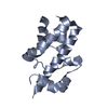
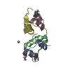
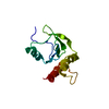
- PDB-3ohw: X-Ray Structure of Phycobilisome LCM core-membrane linker polypep... -
+ Open data

- Basic information
| Entry | Database: PDB / ID: 3ohw | ||||||
|---|---|---|---|---|---|---|---|
| Title | X-Ray Structure of Phycobilisome LCM core-membrane linker polypeptide (fragment 721-860) from Synechocystis sp. PCC 6803, Northeast Structural Genomics Consortium Target SgR209E | ||||||
 Components Components | Phycobilisome LCM core-membrane linker polypeptide | ||||||
 Keywords Keywords |  PROTEIN BINDING / Structural Genomics / PSI-Biology / Protein Structure Initiative / Northeast Structural Genomics Consortium / NESG PROTEIN BINDING / Structural Genomics / PSI-Biology / Protein Structure Initiative / Northeast Structural Genomics Consortium / NESG | ||||||
| Function / homology |  Function and homology informationLyases / phycobilisome / plasma membrane-derived thylakoid membrane / photosynthesis / lyase activity Function and homology informationLyases / phycobilisome / plasma membrane-derived thylakoid membrane / photosynthesis / lyase activitySimilarity search - Function | ||||||
| Biological species |  | ||||||
| Method | X-RAY DIFFRACTION / SYNCHROTRON / SAD / Resolution: 2.7 Å | ||||||
 Authors Authors | Kuzin, A. / Su, M. / Lew, S. / Vorobiev, S.M. / Patel, P. / Xiao, R. / Ciccosanti, C. / Lee, D. / Everett, J.K. / Nair, R. ...Kuzin, A. / Su, M. / Lew, S. / Vorobiev, S.M. / Patel, P. / Xiao, R. / Ciccosanti, C. / Lee, D. / Everett, J.K. / Nair, R. / Acton, T.B. / Rost, B. / Montelione, G.T. / Hunt, J.F. / Tong, L. / Northeast Structural Genomics Consortium (NESG) | ||||||
 Citation Citation | Journal: To be Published Title: Northeast Structural Genomics Consortium Target SgR209E Authors: Kuzin, A. / Su, M. / Lew, S. / Vorobiev, S.M. / Patel, P. / Xiao, R. / Ciccosanti, C. / Lee, D. / Everett, J.K. / Nair, R. / Acton, T.B. / Rost, B. / Montelione, G.T. / Hunt, J.F. / Tong, L. | ||||||
| History |
|
- Structure visualization
| Structure viewer | Molecule: MolmilJmol/JSmol |
|---|
- Downloads & links
-Download
| PDBx/mmCIF format | 3ohw.cif.gz | 107.6 KB | Display | PDBx/mmCIF format |
|---|---|---|---|---|
| PDB format | pdb3ohw.ent.gz | 88.5 KB | Display | PDB format |
| PDBx/mmJSON format | 3ohw.json.gz | Tree view | PDBx/mmJSON format | |
| Others |  Other downloads Other downloads |
-Validation report
| Arichive directory | https://data.pdbj.org/pub/pdb/validation_reports/oh/3ohwftp://data.pdbj.org/pub/pdb/validation_reports/oh/3ohw | HTTPS FTP |
|---|
-Related structure data
| Related structure data | |
|---|---|
| Similar structure data | |
| Other databases |


-Links
 PDBj
PDBj- Assembly
| Deposited unit | 
| ||||||||
|---|---|---|---|---|---|---|---|---|---|
| 1 | 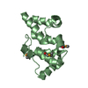
| ||||||||
| 2 | 
| ||||||||
| Unit cell |
|
-Components
| #1: Protein | Mass: 17311.059 Da / Num. of mol.: 2 / Fragment: sequence database residues 711-858 Source method: isolated from a genetically manipulated source Source: (gene. exp.) Escherichia coli (E. coli) / Strain (production host): BL21(DE3)+ Magic / References: UniProt: Q55544 |
|---|
-Experimental details
-Experiment
| Experiment | Method: X-RAY DIFFRACTION / Number of used crystals: 1 |
|---|
- Sample preparation
| Crystal | Density Matthews: 2.15 Å3/Da / Density % sol: 42.66 % |
|---|---|
| Crystal grow | Temperature: 297 K / Method: macrobatch under oik / pH: 7 Details: Protein solution: 100mM NaCl, 5mM DTT, 0.02% NaN3, 10mM Tris-HCl (pH 7.5), Reservoir solution:tryptone 1% (w/v), HEPES sodium 0.05M, PEG3350 20% (w/v), macrobatch under oik, temperature 297K |
-Data collection
| Diffraction | Mean temperature: 100 K |
|---|---|
| Diffraction source | Source: SYNCHROTRON / Site: NSLS  / Beamline: X4A / Wavelength: 0.979 Å / Beamline: X4A / Wavelength: 0.979 Å |
| Detector | Type: ADSC QUANTUM 4r / Detector: CCD / Date: Aug 10, 2010 |
| Radiation | Monochromator: Si 111 CHANNEL / Protocol: SINGLE WAVELENGTH / Monochromatic (M) / Laue (L): M / Scattering type: x-ray |
| Radiation wavelength | Wavelength: 0.979 Å / Relative weight: 1 |
| Reflection | Resolution: 2.7→30 Å / Num. obs: 15697 / % possible obs: 98 % / Observed criterion σ(I): -3 / Redundancy: 3.4 % / Biso Wilson estimate: 26.93 Å2 / Rmerge(I) obs: 0.112 / Net I/σ(I): 10.9 |
- Processing
| Software |
| |||||||||||||||||||||||||||||||||||||||||||||||||
|---|---|---|---|---|---|---|---|---|---|---|---|---|---|---|---|---|---|---|---|---|---|---|---|---|---|---|---|---|---|---|---|---|---|---|---|---|---|---|---|---|---|---|---|---|---|---|---|---|---|---|
| Refinement | Method to determine structure: SAD / Resolution: 2.7→27.753 Å / Occupancy max: 1 / Occupancy min: 0.5 / FOM work R set: 0.816 / SU ML: 0.39 / σ(F): 5.81 / Phase error: 25.62 / Stereochemistry target values: ML
| |||||||||||||||||||||||||||||||||||||||||||||||||
| Solvent computation | Shrinkage radii: 0.72 Å / VDW probe radii: 1 Å / Solvent model: FLAT BULK SOLVENT MODEL / Bsol: 17.948 Å2 / ksol: 0.333 e/Å3 | |||||||||||||||||||||||||||||||||||||||||||||||||
| Displacement parameters | Biso max: 186.68 Å2 / Biso mean: 27.447 Å2 / Biso min: 2.05 Å2
| |||||||||||||||||||||||||||||||||||||||||||||||||
| Refinement step | Cycle: LAST / Resolution: 2.7→27.753 Å
| |||||||||||||||||||||||||||||||||||||||||||||||||
| Refine LS restraints |
| |||||||||||||||||||||||||||||||||||||||||||||||||
| LS refinement shell | Refine-ID: X-RAY DIFFRACTION / Total num. of bins used: 6
| |||||||||||||||||||||||||||||||||||||||||||||||||
| Refinement TLS params. | Method: refined / Origin x: 13.6927 Å / Origin y: 1.4943 Å / Origin z: 17.0359 Å
| |||||||||||||||||||||||||||||||||||||||||||||||||
| Refinement TLS group |
|
