 Movie
Movie Controller

[English] 日本語
 Yorodumi
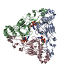
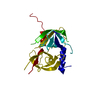
- PDB-3nwu: Substrate induced remodeling of the active site regulates HtrA1 a... -
+ Open data

- Basic information
| Entry | Database: PDB / ID: 3nwu | ||||||
|---|---|---|---|---|---|---|---|
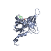
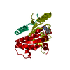
| Title | Substrate induced remodeling of the active site regulates HtrA1 activity | ||||||
 Components Components | Serine protease HTRA1 | ||||||
 Keywords Keywords |  HYDROLASE / serine protease / DegP / HtrA / protease HYDROLASE / serine protease / DegP / HtrA / protease | ||||||
| Function / homology |  Function and homology information Function and homology informationchorionic trophoblast cell differentiation / programmed cell death / growth factor binding / negative regulation of BMP signaling pathway / Hydrolases; Acting on peptide bonds (peptidases); Serine endopeptidases / serine-type peptidase activity / Degradation of the extracellular matrix / molecular function activator activity / placenta development / negative regulation of transforming growth factor beta receptor signaling pathway ...chorionic trophoblast cell differentiation / programmed cell death / growth factor binding / negative regulation of BMP signaling pathway / Hydrolases; Acting on peptide bonds (peptidases); Serine endopeptidases / serine-type peptidase activity / Degradation of the extracellular matrix / molecular function activator activity / placenta development / negative regulation of transforming growth factor beta receptor signaling pathway / collagen-containing extracellular matrix / positive regulation of apoptotic process / serine-type endopeptidase activity / proteolysis / extracellular space / extracellular exosome / extracellular region / identical protein binding / plasma membrane / cytosolSimilarity search - Function | ||||||
| Biological species |  Homo sapiens (human) Homo sapiens (human) | ||||||
| Method | X-RAY DIFFRACTION / SYNCHROTRON / MOLECULAR REPLACEMENT / Resolution: 3.2 Å | ||||||
 Authors Authors | Truebestein, L. / Tennstaedt, A. / Hauske, P. / Krojer, T. / Kaiser, M. / Clausen, T. / Ehrmann, M. | ||||||
 Citation Citation | Journal: Nat.Struct.Mol.Biol. / Year: 2011 Title: Substrate-induced remodeling of the active site regulates human HTRA1 activity. Authors: Truebestein, L. / Tennstaedt, A. / Monig, T. / Krojer, T. / Canellas, F. / Kaiser, M. / Clausen, T. / Ehrmann, M. | ||||||
| History |
|
- Structure visualization
| Structure viewer | Molecule: MolmilJmol/JSmol |
|---|
- Downloads & links
-Download
| PDBx/mmCIF format | 3nwu.cif.gz | 114.9 KB | Display | PDBx/mmCIF format |
|---|---|---|---|---|
| PDB format | pdb3nwu.ent.gz | 90.5 KB | Display | PDB format |
| PDBx/mmJSON format | 3nwu.json.gz | Tree view | PDBx/mmJSON format | |
| Others |  Other downloads Other downloads |
-Validation report
| Arichive directory | https://data.pdbj.org/pub/pdb/validation_reports/nw/3nwuftp://data.pdbj.org/pub/pdb/validation_reports/nw/3nwu | HTTPS FTP |
|---|
-Related structure data


-Links
 PDBj
PDBj


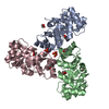
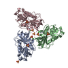
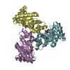
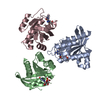
- Assembly
| Deposited unit | 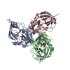
| ||||||||||||||||||||||||
|---|---|---|---|---|---|---|---|---|---|---|---|---|---|---|---|---|---|---|---|---|---|---|---|---|---|
| 1 |
| ||||||||||||||||||||||||
| 2 | 
| ||||||||||||||||||||||||
| Unit cell |
| ||||||||||||||||||||||||
| Noncrystallographic symmetry (NCS) | NCS domain:
|
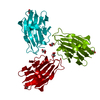
-Components
| #1: Protein | Mass: 24876.395 Da / Num. of mol.: 3 / Fragment: HtrA1 protease domain (unp residues 158-375) Source method: isolated from a genetically manipulated source Source: (gene. exp.) Homo sapiens (human) / Gene: HTRA, HTRA1, PRSS11 / Production host:  Escherichia coli (E. coli) Escherichia coli (E. coli)References: UniProt: Q92743, Hydrolases; Acting on peptide bonds (peptidases); Serine endopeptidases#2: Chemical | Sulfate  Mass: 96.063 Da / Num. of mol.: 3 / Source method: obtained synthetically / Formula: SO4 Mass: 96.063 Da / Num. of mol.: 3 / Source method: obtained synthetically / Formula: SO4 |
|---|
-Experimental details
-Experiment
| Experiment | Method: X-RAY DIFFRACTION / Number of used crystals: 1 |
|---|
- Sample preparation
| Crystal | Density Matthews: 3.55 Å3/Da / Density % sol: 65.38 % |
|---|---|
| Crystal grow | Temperature: 292 K / pH: 4.6 Details: 2.0M Ammonium sulfate, 0.1M Sodium acetate, pH 4.6, VAPOR DIFFUSION, SITTING DROP, temperature 292K |
-Data collection
| Diffraction | Mean temperature: 100 K |
|---|---|
| Diffraction source | Source: SYNCHROTRON / Site: ESRF  / Beamline: ID14-4 / Wavelength: 1.0809 Å / Beamline: ID14-4 / Wavelength: 1.0809 Å |
| Detector | Type: ADSC QUANTUM 315 / Detector: CCD / Date: Dec 2, 2007 |
| Radiation | Protocol: SINGLE WAVELENGTH / Monochromatic (M) / Laue (L): M / Scattering type: x-ray |
| Radiation wavelength | Wavelength: 1.0809 Å / Relative weight: 1 |
| Reflection | Resolution: 3.2→19.9 Å / Num. obs: 17660 / % possible obs: 97.1 % / Observed criterion σ(I): -3 / Redundancy: 3.7 % / Biso Wilson estimate: 58.3 Å2 / Rmerge(I) obs: 0.099 / Net I/σ(I): 15.2 |
| Reflection shell | Resolution: 3.2→3.31 Å / Redundancy: 3.6 % / Rmerge(I) obs: 0.469 / Mean I/σ(I) obs: 2.9 / % possible all: 97.8 |
- Processing
| Software |
| |||||||||||||||||||||||||||||||||||||||||||||||||||||||||||||||||||||||||||||||||||||||||||||||||||||||||||||||||||||||||||||||||||||||||||||||||||||||||||||||||||||||||||||||
|---|---|---|---|---|---|---|---|---|---|---|---|---|---|---|---|---|---|---|---|---|---|---|---|---|---|---|---|---|---|---|---|---|---|---|---|---|---|---|---|---|---|---|---|---|---|---|---|---|---|---|---|---|---|---|---|---|---|---|---|---|---|---|---|---|---|---|---|---|---|---|---|---|---|---|---|---|---|---|---|---|---|---|---|---|---|---|---|---|---|---|---|---|---|---|---|---|---|---|---|---|---|---|---|---|---|---|---|---|---|---|---|---|---|---|---|---|---|---|---|---|---|---|---|---|---|---|---|---|---|---|---|---|---|---|---|---|---|---|---|---|---|---|---|---|---|---|---|---|---|---|---|---|---|---|---|---|---|---|---|---|---|---|---|---|---|---|---|---|---|---|---|---|---|---|---|---|
| Refinement | Method to determine structure: MOLECULAR REPLACEMENT / Resolution: 3.2→19.9 Å / Cor.coef. Fo:Fc: 0.949 / Cor.coef. Fo:Fc free: 0.935 / SU B: 13.467 / SU ML: 0.233 / Cross valid method: THROUGHOUT / ESU R Free: 0.343 / Stereochemistry target values: MAXIMUM LIKELIHOOD / Details: HYDROGENS HAVE BEEN USED IF PRESENT IN THE INPUT
| |||||||||||||||||||||||||||||||||||||||||||||||||||||||||||||||||||||||||||||||||||||||||||||||||||||||||||||||||||||||||||||||||||||||||||||||||||||||||||||||||||||||||||||||
| Solvent computation | Ion probe radii: 0.8 Å / Shrinkage radii: 0.8 Å / VDW probe radii: 1.2 Å / Solvent model: MASK | |||||||||||||||||||||||||||||||||||||||||||||||||||||||||||||||||||||||||||||||||||||||||||||||||||||||||||||||||||||||||||||||||||||||||||||||||||||||||||||||||||||||||||||||
| Displacement parameters | Biso mean: 49.2 Å2
| |||||||||||||||||||||||||||||||||||||||||||||||||||||||||||||||||||||||||||||||||||||||||||||||||||||||||||||||||||||||||||||||||||||||||||||||||||||||||||||||||||||||||||||||
| Refinement step | Cycle: LAST / Resolution: 3.2→19.9 Å
| |||||||||||||||||||||||||||||||||||||||||||||||||||||||||||||||||||||||||||||||||||||||||||||||||||||||||||||||||||||||||||||||||||||||||||||||||||||||||||||||||||||||||||||||
| Refine LS restraints |
| |||||||||||||||||||||||||||||||||||||||||||||||||||||||||||||||||||||||||||||||||||||||||||||||||||||||||||||||||||||||||||||||||||||||||||||||||||||||||||||||||||||||||||||||
| Refine LS restraints NCS | Refine-ID: X-RAY DIFFRACTION
| |||||||||||||||||||||||||||||||||||||||||||||||||||||||||||||||||||||||||||||||||||||||||||||||||||||||||||||||||||||||||||||||||||||||||||||||||||||||||||||||||||||||||||||||
| LS refinement shell | Resolution: 3.2→3.28 Å / Total num. of bins used: 20
|
