 Movie
Movie Controller

[English] 日本語
 Yorodumi
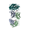
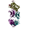
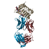
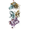
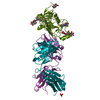
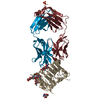
- PDB-3nps: Crystal structure of membrane-type serine protease 1 (MT-SP1) in ... -
+ Open data

- Basic information
| Entry | Database: PDB / ID: 3nps | ||||||
|---|---|---|---|---|---|---|---|
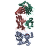
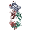
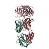
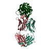
| Title | Crystal structure of membrane-type serine protease 1 (MT-SP1) in complex with the Fab Inhibitor S4 | ||||||
 Components Components |
| ||||||
 Keywords Keywords | HYDROLASE/IMMUNE SYSTEM /  hydrolase / Antibody-Protease Complex / Protein-Protein Complex / Enzyme-Inhibitor Complex / Disease mutation / Glycoprotein / Membrane / Serine protease / Signal-anchor / Transmembrane / HYDROLASE - IMMUNE SYSTEM complex / HYDROLASE-IMMUNE SYSTEM complex hydrolase / Antibody-Protease Complex / Protein-Protein Complex / Enzyme-Inhibitor Complex / Disease mutation / Glycoprotein / Membrane / Serine protease / Signal-anchor / Transmembrane / HYDROLASE - IMMUNE SYSTEM complex / HYDROLASE-IMMUNE SYSTEM complex | ||||||
| Function / homology |  Function and homology informationmatriptase / epithelial cell morphogenesis involved in placental branching / acrosome reaction / Formation of the cornified envelope / keratinocyte differentiation / serine-type peptidase activity / neural tube closure / protein catabolic process / basolateral plasma membrane / external side of plasma membrane ...matriptase / epithelial cell morphogenesis involved in placental branching / acrosome reaction / Formation of the cornified envelope / keratinocyte differentiation / serine-type peptidase activity / neural tube closure / protein catabolic process / basolateral plasma membrane / external side of plasma membrane / serine-type endopeptidase activity / proteolysis / extracellular space / plasma membrane Function and homology informationmatriptase / epithelial cell morphogenesis involved in placental branching / acrosome reaction / Formation of the cornified envelope / keratinocyte differentiation / serine-type peptidase activity / neural tube closure / protein catabolic process / basolateral plasma membrane / external side of plasma membrane ...matriptase / epithelial cell morphogenesis involved in placental branching / acrosome reaction / Formation of the cornified envelope / keratinocyte differentiation / serine-type peptidase activity / neural tube closure / protein catabolic process / basolateral plasma membrane / external side of plasma membrane / serine-type endopeptidase activity / proteolysis / extracellular space / plasma membraneSimilarity search - Function | ||||||
| Biological species |  Homo sapiens (human) Homo sapiens (human) | ||||||
| Method | X-RAY DIFFRACTION / SYNCHROTRON / MOLECULAR REPLACEMENT / molecular replacement / Resolution: 1.5 Å | ||||||
 Authors Authors | Baharuddin, A. | ||||||
 Citation Citation | Journal: J.Mol.Biol. / Year: 2012 Title: A reverse binding motif that contributes to specific protease inhibition by antibodies. Authors: Schneider, E.L. / Lee, M.S. / Baharuddin, A. / Goetz, D.H. / Farady, C.J. / Ward, M. / Wang, C.I. / Craik, C.S. | ||||||
| History |
|
- Structure visualization
| Structure viewer | Molecule: MolmilJmol/JSmol |
|---|
- Downloads & links
-Download
| PDBx/mmCIF format | 3nps.cif.gz | 280.4 KB | Display | PDBx/mmCIF format |
|---|---|---|---|---|
| PDB format | pdb3nps.ent.gz | 225.3 KB | Display | PDB format |
| PDBx/mmJSON format | 3nps.json.gz | Tree view | PDBx/mmJSON format | |
| Others |  Other downloads Other downloads |
-Validation report
| Arichive directory | https://data.pdbj.org/pub/pdb/validation_reports/np/3npsftp://data.pdbj.org/pub/pdb/validation_reports/np/3nps | HTTPS FTP |
|---|
-Related structure data
| Related structure data | 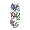 3so3C  2jb5S 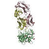 3bn9S 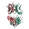 3kdmS S: Starting model for refinement C: citing same article ( |
|---|---|
| Similar structure data |


-Links
 PDBj
PDBj


- Assembly
| Deposited unit | 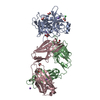
| ||||||||
|---|---|---|---|---|---|---|---|---|---|
| 1 |
| ||||||||
| Unit cell |
|
-Components
-Protein , 1 types, 1 molecules A
| #1: Protein | Mass: 26447.689 Da / Num. of mol.: 1 / Fragment: PEPTIDASE S1 DOMAIN (unp residues 615-855) / Mutation: C122S Source method: isolated from a genetically manipulated source Source: (gene. exp.) Homo sapiens (human) / Gene: PRSS14, SNC19, ST14, TADG15 / Plasmid: pQE30 / Production host:  Escherichia coli (E. coli) / Strain (production host): BL21(DE3) / References: UniProt: Q9Y5Y6, matriptase Escherichia coli (E. coli) / Strain (production host): BL21(DE3) / References: UniProt: Q9Y5Y6, matriptase |
|---|
-Antibody , 2 types, 2 molecules BC
| #2: Antibody | Mass: 24134.084 Da / Num. of mol.: 1 Source method: isolated from a genetically manipulated source Source: (gene. exp.) Homo sapiens (human) / Plasmid: pMx7FH / Production host: Escherichia coli (E. coli) |
|---|---|
| #3: Antibody | Mass: 22731.070 Da / Num. of mol.: 1 Source method: isolated from a genetically manipulated source Source: (gene. exp.) Homo sapiens (human) / Plasmid: pMx7FH / Production host: Escherichia coli (E. coli) |
-Non-polymers , 4 types, 692 molecules 


| #4: Chemical | ChemComp-EDO / Ethylene glycol Mass: 62.068 Da / Num. of mol.: 10 / Source method: obtained synthetically / Formula: C2H6O2 Mass: 62.068 Da / Num. of mol.: 10 / Source method: obtained synthetically / Formula: C2H6O2#5: Chemical | ChemComp-NA /  Mass: 22.990 Da / Num. of mol.: 5 / Source method: obtained synthetically / Formula: Na Mass: 22.990 Da / Num. of mol.: 5 / Source method: obtained synthetically / Formula: Na#6: Chemical | ChemComp-CL / | Chloride Mass: 35.453 Da / Num. of mol.: 1 / Source method: obtained synthetically / Formula: Cl Mass: 35.453 Da / Num. of mol.: 1 / Source method: obtained synthetically / Formula: Cl#7: Water | ChemComp-HOH / | WaterMass: 18.015 Da / Num. of mol.: 676 / Source method: isolated from a natural source / Formula: H2O |
|---|
-Experimental details
-Experiment
| Experiment | Method: X-RAY DIFFRACTION / Number of used crystals: 1 |
|---|
- Sample preparation
| Crystal | Density Matthews: 2.27 Å3/Da / Density % sol: 45.9 % |
|---|---|
| Crystal grow | Temperature: 298 K Details: 50 mM Tris, 100 mM NaCl, 5% glycerol, no buffer was added for crystallization, VAPOR DIFFUSION, HANGING DROP, temperature 298K |
-Data collection
| Diffraction | Mean temperature: 100 K |
|---|---|
| Diffraction source | Source: SYNCHROTRON / Site: ALS  / Beamline: 8.3.1 / Wavelength: 1 / Beamline: 8.3.1 / Wavelength: 1 |
| Detector | Type: ADSC QUANTUM 210 / Detector: CCD / Date: Jan 1, 2010 / Details: MIRRORS |
| Radiation | Monochromator: KOHZU DOUBLE CRYSTAL SI (111) / Protocol: SINGLE WAVELENGTH / Monochromatic (M) / Laue (L): M / Scattering type: x-ray |
| Radiation wavelength | Wavelength: 1 Å / Relative weight: 1 |
| Reflection | Resolution: 1.4→43.393 Å / Num. obs: 88531 / % possible obs: 84.5 % / Observed criterion σ(I): 1 / Redundancy: 3.7 % / Biso Wilson estimate: 19.6 Å2 / Rmerge(I) obs: 0.058 / Net I/σ(I): 16 |
| Reflection shell | Resolution: 1.4→1.46 Å / Redundancy: 3.1 % / Rmerge(I) obs: 0.176 / Mean I/σ(I) obs: 8 / % possible all: 80 |
-Phasing
| Phasing | Method: molecular replacement | |||||||||
|---|---|---|---|---|---|---|---|---|---|---|
| Phasing MR | Rfactor: 46.15 / Model details: Phaser MODE: MR_AUTO
|
- Processing
| Software |
| ||||||||||||||||||||||||||||||||||||||||||||||||||||||||||||||||||||||||||||||||||||||||||||||||||||||||||||||||||||||||||||||||||||||||||||||||||||||||||||||||||||||||||
|---|---|---|---|---|---|---|---|---|---|---|---|---|---|---|---|---|---|---|---|---|---|---|---|---|---|---|---|---|---|---|---|---|---|---|---|---|---|---|---|---|---|---|---|---|---|---|---|---|---|---|---|---|---|---|---|---|---|---|---|---|---|---|---|---|---|---|---|---|---|---|---|---|---|---|---|---|---|---|---|---|---|---|---|---|---|---|---|---|---|---|---|---|---|---|---|---|---|---|---|---|---|---|---|---|---|---|---|---|---|---|---|---|---|---|---|---|---|---|---|---|---|---|---|---|---|---|---|---|---|---|---|---|---|---|---|---|---|---|---|---|---|---|---|---|---|---|---|---|---|---|---|---|---|---|---|---|---|---|---|---|---|---|---|---|---|---|---|---|---|---|---|
| Refinement | Method to determine structure: MOLECULAR REPLACEMENT Starting model: PDB ENTRIES: 3BN9, 2JB5, 3KDM Resolution: 1.5→28.64 Å / Cor.coef. Fo:Fc: 0.966 / Cor.coef. Fo:Fc free: 0.951 / Occupancy max: 1 / Occupancy min: 0 / SU B: 1.661 / SU ML: 0.062 / Cross valid method: THROUGHOUT / σ(F): 0 / ESU R Free: 0.098 / Stereochemistry target values: MAXIMUM LIKELIHOOD / Details: U VALUES : REFINED INDIVIDUALLY
| ||||||||||||||||||||||||||||||||||||||||||||||||||||||||||||||||||||||||||||||||||||||||||||||||||||||||||||||||||||||||||||||||||||||||||||||||||||||||||||||||||||||||||
| Solvent computation | Ion probe radii: 0.8 Å / Shrinkage radii: 0.8 Å / VDW probe radii: 1.2 Å / Solvent model: BABINET MODEL WITH MASK | ||||||||||||||||||||||||||||||||||||||||||||||||||||||||||||||||||||||||||||||||||||||||||||||||||||||||||||||||||||||||||||||||||||||||||||||||||||||||||||||||||||||||||
| Displacement parameters | Biso mean: 23.94 Å2
| ||||||||||||||||||||||||||||||||||||||||||||||||||||||||||||||||||||||||||||||||||||||||||||||||||||||||||||||||||||||||||||||||||||||||||||||||||||||||||||||||||||||||||
| Refine analyze |
| ||||||||||||||||||||||||||||||||||||||||||||||||||||||||||||||||||||||||||||||||||||||||||||||||||||||||||||||||||||||||||||||||||||||||||||||||||||||||||||||||||||||||||
| Refinement step | Cycle: LAST / Resolution: 1.5→28.64 Å
| ||||||||||||||||||||||||||||||||||||||||||||||||||||||||||||||||||||||||||||||||||||||||||||||||||||||||||||||||||||||||||||||||||||||||||||||||||||||||||||||||||||||||||
| Refine LS restraints |
| ||||||||||||||||||||||||||||||||||||||||||||||||||||||||||||||||||||||||||||||||||||||||||||||||||||||||||||||||||||||||||||||||||||||||||||||||||||||||||||||||||||||||||
| LS refinement shell | Resolution: 1.5→1.54 Å / Total num. of bins used: 20
| ||||||||||||||||||||||||||||||||||||||||||||||||||||||||||||||||||||||||||||||||||||||||||||||||||||||||||||||||||||||||||||||||||||||||||||||||||||||||||||||||||||||||||
| Refinement TLS params. | Method: refined / Refine-ID: X-RAY DIFFRACTION
| ||||||||||||||||||||||||||||||||||||||||||||||||||||||||||||||||||||||||||||||||||||||||||||||||||||||||||||||||||||||||||||||||||||||||||||||||||||||||||||||||||||||||||
| Refinement TLS group |
|
