 Movie
Movie Controller

[English] 日本語
 Yorodumi
- PDB-3kdm: Crystal Structure of Human Anti-steroid Fab 5F2 in Complex with T... -
+ Open data

- Basic information
| Entry | Database: PDB / ID: 3kdm | |||||||||
|---|---|---|---|---|---|---|---|---|---|---|
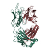
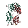
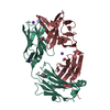
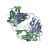
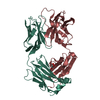
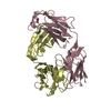
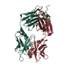
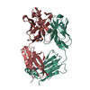
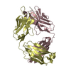
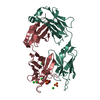
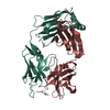
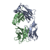
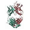
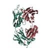
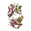
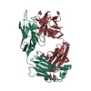
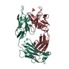
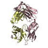
| Title | Crystal Structure of Human Anti-steroid Fab 5F2 in Complex with Testosterone | |||||||||
 Components Components |
| |||||||||
 Keywords Keywords |  IMMUNE SYSTEM / Antibody / Immunoglobulin Fab fragment / Anti-steroid IMMUNE SYSTEM / Antibody / Immunoglobulin Fab fragment / Anti-steroid | |||||||||
| Function / homology | Immunoglobulins / Immunoglobulin-like / Sandwich / Mainly Beta / TESTOSTERONE Function and homology information Function and homology information | |||||||||
| Biological species |  Homo sapiens (human) Homo sapiens (human) | |||||||||
| Method | X-RAY DIFFRACTION / SYNCHROTRON / MOLECULAR REPLACEMENT / Resolution: 1.5 Å | |||||||||
 Authors Authors | Niemi, M.H. / Rouvinen, J. | |||||||||
 Citation Citation | Journal: J.Mol.Recognit. / Year: 2010 Title: The testosterone binding mechanism of an antibody derived from a naive human scFv library Authors: Niemi, M.H. / Takkinen, K. / Amundsen, L.K. / Soderlund, H. / Rouvinen, J. / Hoyhtya, M. | |||||||||
| History |
|
- Structure visualization
| Structure viewer | Molecule: MolmilJmol/JSmol |
|---|
- Downloads & links
-Download
| PDBx/mmCIF format | 3kdm.cif.gz | 195.9 KB | Display | PDBx/mmCIF format |
|---|---|---|---|---|
| PDB format | pdb3kdm.ent.gz | 153.3 KB | Display | PDB format |
| PDBx/mmJSON format | 3kdm.json.gz | Tree view | PDBx/mmJSON format | |
| Others |  Other downloads Other downloads |
-Validation report
| Arichive directory | https://data.pdbj.org/pub/pdb/validation_reports/kd/3kdmftp://data.pdbj.org/pub/pdb/validation_reports/kd/3kdm | HTTPS FTP |
|---|
-Related structure data
| Related structure data |  1mhpS S: Starting model for refinement |
|---|---|
| Similar structure data |


-Links
 PDBj
PDBj


- Assembly
| Deposited unit | 
| ||||||||
|---|---|---|---|---|---|---|---|---|---|
| 1 | 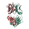
| ||||||||
| 2 | 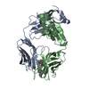
| ||||||||
| Unit cell |
|
-Components
| #1: Antibody | Mass: 23373.709 Da / Num. of mol.: 2 Source method: isolated from a genetically manipulated source Source: (gene. exp.) Homo sapiens (human) / Production host:  Escherichia coli (E. coli) / Strain (production host): RV308 Escherichia coli (E. coli) / Strain (production host): RV308#2: Antibody | Mass: 23726.475 Da / Num. of mol.: 2 Source method: isolated from a genetically manipulated source Source: (gene. exp.) Homo sapiens (human) / Production host: Escherichia coli (E. coli) / Strain (production host): RV308#3: Chemical | Testosterone  Mass: 288.424 Da / Num. of mol.: 2 / Source method: obtained synthetically / Formula: C19H28O2 / Comment: hormone*YM Mass: 288.424 Da / Num. of mol.: 2 / Source method: obtained synthetically / Formula: C19H28O2 / Comment: hormone*YM#4: Water | ChemComp-HOH / | Water Mass: 18.015 Da / Num. of mol.: 919 / Source method: isolated from a natural source / Formula: H2O Mass: 18.015 Da / Num. of mol.: 919 / Source method: isolated from a natural source / Formula: H2O |
|---|
-Experimental details
-Experiment
| Experiment | Method: X-RAY DIFFRACTION / Number of used crystals: 1 |
|---|
- Sample preparation
| Crystal | Density Matthews: 2.79 Å3/Da / Density % sol: 55.87 % |
|---|---|
| Crystal grow | Temperature: 293 K / Method: vapor diffusion, hanging drop / pH: 4.7 Details: 12% PEG 3350, 0.1M sodium citrate, pH 4.7, VAPOR DIFFUSION, HANGING DROP, temperature 293K |
-Data collection
| Diffraction | Mean temperature: 100 K |
|---|---|
| Diffraction source | Source: SYNCHROTRON / Site: ESRF  / Beamline: ID29 / Wavelength: 1.0091 Å / Beamline: ID29 / Wavelength: 1.0091 Å |
| Detector | Type: ADSC QUANTUM 315r / Detector: CCD / Date: Apr 14, 2007 |
| Radiation | Protocol: SINGLE WAVELENGTH / Monochromatic (M) / Laue (L): M / Scattering type: x-ray |
| Radiation wavelength | Wavelength: 1.0091 Å / Relative weight: 1 |
| Reflection twin | Operator: -h,-l,-k / Fraction: 0.501 |
| Reflection | Resolution: 1.5→50 Å / Num. all: 163180 / Num. obs: 151891 / % possible obs: 93.1 % / Observed criterion σ(I): 2 / Redundancy: 2 % / Biso Wilson estimate: 23.8 Å2 / Rmerge(I) obs: 0.033 |
| Reflection shell | Resolution: 1.5→1.6 Å / Redundancy: 2 % / Rmerge(I) obs: 0.321 / Mean I/σ(I) obs: 2.47 / % possible all: 91 |
- Processing
| Software |
| ||||||||||||||||||||||||||||||||||||||||||||||||||||||||||||||||||||||||||||||||||||||||||||||||||||||||||||||||||||||||||||||
|---|---|---|---|---|---|---|---|---|---|---|---|---|---|---|---|---|---|---|---|---|---|---|---|---|---|---|---|---|---|---|---|---|---|---|---|---|---|---|---|---|---|---|---|---|---|---|---|---|---|---|---|---|---|---|---|---|---|---|---|---|---|---|---|---|---|---|---|---|---|---|---|---|---|---|---|---|---|---|---|---|---|---|---|---|---|---|---|---|---|---|---|---|---|---|---|---|---|---|---|---|---|---|---|---|---|---|---|---|---|---|---|---|---|---|---|---|---|---|---|---|---|---|---|---|---|---|---|
| Refinement | Method to determine structure: MOLECULAR REPLACEMENT Starting model: PDB ENTRY 1MHP Resolution: 1.5→48.25 Å / Occupancy max: 1 / Occupancy min: 0.5 / FOM work R set: 0.832 / σ(F): 1.99
| ||||||||||||||||||||||||||||||||||||||||||||||||||||||||||||||||||||||||||||||||||||||||||||||||||||||||||||||||||||||||||||||
| Solvent computation | Shrinkage radii: 0.9 Å / VDW probe radii: 1.11 Å / Solvent model: FLAT BULK SOLVENT MODEL / Bsol: 50.896 Å2 / ksol: 0.37 e/Å3 | ||||||||||||||||||||||||||||||||||||||||||||||||||||||||||||||||||||||||||||||||||||||||||||||||||||||||||||||||||||||||||||||
| Displacement parameters | Biso max: 59.06 Å2 / Biso mean: 20.116 Å2 / Biso min: 10.81 Å2
| ||||||||||||||||||||||||||||||||||||||||||||||||||||||||||||||||||||||||||||||||||||||||||||||||||||||||||||||||||||||||||||||
| Refinement step | Cycle: LAST / Resolution: 1.5→48.25 Å
| ||||||||||||||||||||||||||||||||||||||||||||||||||||||||||||||||||||||||||||||||||||||||||||||||||||||||||||||||||||||||||||||
| Refine LS restraints |
| ||||||||||||||||||||||||||||||||||||||||||||||||||||||||||||||||||||||||||||||||||||||||||||||||||||||||||||||||||||||||||||||
| LS refinement shell | Refine-ID: X-RAY DIFFRACTION / Total num. of bins used: 20 / % reflection obs: 98 %
|
