 Movie
Movie Controller

[English] 日本語
 Yorodumi
- PDB-3ma2: Complex membrane type-1 matrix metalloproteinase (MT1-MMP) with t... -
+ Open data

- Basic information
| Entry | Database: PDB / ID: 3ma2 | ||||||
|---|---|---|---|---|---|---|---|
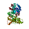
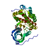
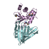
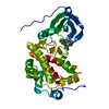
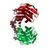
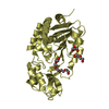
| Title | Complex membrane type-1 matrix metalloproteinase (MT1-MMP) with tissue inhibitor of metalloproteinase-1 (TIMP-1) | ||||||
 Components Components |
| ||||||
 Keywords Keywords | HYDROLASE/HYDROLASE INHIBITOR / Protein - protein complex / Cleavage on pair of basic residues /  Disulfide bond / Membrane / Metal-binding / Metalloprotease / Protease / Transmembrane / Zymogen / Erythrocyte maturation / Glycoprotein / Metalloenzyme inhibitor / Metalloprotease inhibitor / Secreted / HYDROLASE-HYDROLASE INHIBITOR complex Disulfide bond / Membrane / Metal-binding / Metalloprotease / Protease / Transmembrane / Zymogen / Erythrocyte maturation / Glycoprotein / Metalloenzyme inhibitor / Metalloprotease inhibitor / Secreted / HYDROLASE-HYDROLASE INHIBITOR complex | ||||||
| Function / homology |  Function and homology informationmembrane-type matrix metalloproteinase-1 / craniofacial suture morphogenesis / regulation of integrin-mediated signaling pathway / negative regulation of metallopeptidase activity / positive regulation of macrophage migration / macropinosome / peptidase inhibitor activity / head development / negative regulation of trophoblast cell migration / connective tissue replacement involved in inflammatory response wound healing ...membrane-type matrix metalloproteinase-1 / craniofacial suture morphogenesis / regulation of integrin-mediated signaling pathway / negative regulation of metallopeptidase activity / positive regulation of macrophage migration / macropinosome / peptidase inhibitor activity / head development / negative regulation of trophoblast cell migration / connective tissue replacement involved in inflammatory response wound healing / negative regulation of membrane protein ectodomain proteolysis / chondrocyte proliferation / astrocyte cell migration / metalloendopeptidase inhibitor activity / response to odorant / tissue remodeling / cellular response to UV-A / negative regulation of catalytic activity / negative regulation of focal adhesion assembly / positive regulation of protein processing / negative regulation of endopeptidase activity / endochondral ossification / embryonic cranial skeleton morphogenesis / cartilage development / zymogen activation / endothelial cell proliferation / intermediate filament cytoskeleton / positive regulation of B cell differentiation / branching morphogenesis of an epithelial tube / positive regulation of myotube differentiation / negative regulation of Notch signaling pathway / endodermal cell differentiation / Activation of Matrix Metalloproteinases / metalloaminopeptidase activity / Interleukin-10 signaling / Collagen degradation / collagen catabolic process / basement membrane / extracellular matrix disassembly / regulation of protein localization to plasma membrane / response to mechanical stimulus / ovarian follicle development / Degradation of the extracellular matrix / extracellular matrix organization / extracellular matrix / platelet alpha granule lumen / response to hormone / skeletal system development / response to cytokine / cytokine activity / cell motility / Post-translational protein phosphorylation / growth factor activity / lung development / response to organic cyclic compound / protein processing / metalloendopeptidase activity / response to peptide hormone / Golgi lumen / response to estrogen / Regulation of Insulin-like Growth Factor (IGF) transport and uptake by Insulin-like Growth Factor Binding Proteins (IGFBPs) / male gonad development / melanosome / integrin binding / Platelet degranulation / cytoplasmic vesicle / positive regulation of cell growth / Interleukin-4 and Interleukin-13 signaling / angiogenesis / endopeptidase activity / protease binding / response to oxidative stress / response to hypoxia / positive regulation of cell migration / endoplasmic reticulum lumen / serine-type endopeptidase activity / focal adhesion / positive regulation of cell population proliferation / negative regulation of apoptotic process / proteolysis / extracellular space / extracellular exosome / zinc ion binding / extracellular region / nucleus / plasma membrane / cytosol Function and homology informationmembrane-type matrix metalloproteinase-1 / craniofacial suture morphogenesis / regulation of integrin-mediated signaling pathway / negative regulation of metallopeptidase activity / positive regulation of macrophage migration / macropinosome / peptidase inhibitor activity / head development / negative regulation of trophoblast cell migration / connective tissue replacement involved in inflammatory response wound healing ...membrane-type matrix metalloproteinase-1 / craniofacial suture morphogenesis / regulation of integrin-mediated signaling pathway / negative regulation of metallopeptidase activity / positive regulation of macrophage migration / macropinosome / peptidase inhibitor activity / head development / negative regulation of trophoblast cell migration / connective tissue replacement involved in inflammatory response wound healing / negative regulation of membrane protein ectodomain proteolysis / chondrocyte proliferation / astrocyte cell migration / metalloendopeptidase inhibitor activity / response to odorant / tissue remodeling / cellular response to UV-A / negative regulation of catalytic activity / negative regulation of focal adhesion assembly / positive regulation of protein processing / negative regulation of endopeptidase activity / endochondral ossification / embryonic cranial skeleton morphogenesis / cartilage development / zymogen activation / endothelial cell proliferation / intermediate filament cytoskeleton / positive regulation of B cell differentiation / branching morphogenesis of an epithelial tube / positive regulation of myotube differentiation / negative regulation of Notch signaling pathway / endodermal cell differentiation / Activation of Matrix Metalloproteinases / metalloaminopeptidase activity / Interleukin-10 signaling / Collagen degradation / collagen catabolic process / basement membrane / extracellular matrix disassembly / regulation of protein localization to plasma membrane / response to mechanical stimulus / ovarian follicle development / Degradation of the extracellular matrix / extracellular matrix organization / extracellular matrix / platelet alpha granule lumen / response to hormone / skeletal system development / response to cytokine / cytokine activity / cell motility / Post-translational protein phosphorylation / growth factor activity / lung development / response to organic cyclic compound / protein processing / metalloendopeptidase activity / response to peptide hormone / Golgi lumen / response to estrogen / Regulation of Insulin-like Growth Factor (IGF) transport and uptake by Insulin-like Growth Factor Binding Proteins (IGFBPs) / male gonad development / melanosome / integrin binding / Platelet degranulation / cytoplasmic vesicle / positive regulation of cell growth / Interleukin-4 and Interleukin-13 signaling / angiogenesis / endopeptidase activity / protease binding / response to oxidative stress / response to hypoxia / positive regulation of cell migration / endoplasmic reticulum lumen / serine-type endopeptidase activity / focal adhesion / positive regulation of cell population proliferation / negative regulation of apoptotic process / proteolysis / extracellular space / extracellular exosome / zinc ion binding / extracellular region / nucleus / plasma membrane / cytosolSimilarity search - Function | ||||||
| Biological species |  Homo sapiens (human) Homo sapiens (human) | ||||||
| Method | X-RAY DIFFRACTION / MOLECULAR REPLACEMENT / Resolution: 2.05 Å | ||||||
 Authors Authors | Grossman, M. / Tworowski, D. / Dym, O. / Lee, M.-H. / Levy, Y. / Sagi, I. | ||||||
 Citation Citation | Journal: Biochemistry / Year: 2010 Title: The Intrinsic Protein Flexibility of Endogenous Protease Inhibitor TIMP-1 Controls Its Binding Interface and Affects Its Function. Authors: Grossman, M. / Tworowski, D. / Dym, O. / Lee, M.H. / Levy, Y. / Murphy, G. / Sagi, I. | ||||||
| History |
|
- Structure visualization
| Structure viewer | Molecule: MolmilJmol/JSmol |
|---|
- Downloads & links
-Download
| PDBx/mmCIF format | 3ma2.cif.gz | 131.9 KB | Display | PDBx/mmCIF format |
|---|---|---|---|---|
| PDB format | pdb3ma2.ent.gz | 101.3 KB | Display | PDB format |
| PDBx/mmJSON format | 3ma2.json.gz | Tree view | PDBx/mmJSON format | |
| Others |  Other downloads Other downloads |
-Validation report
| Arichive directory | https://data.pdbj.org/pub/pdb/validation_reports/ma/3ma2ftp://data.pdbj.org/pub/pdb/validation_reports/ma/3ma2 | HTTPS FTP |
|---|
-Related structure data
| Related structure data |  1bqqS S: Starting model for refinement |
|---|---|
| Similar structure data |


-Links
 PDBj
PDBj


- Assembly
| Deposited unit | 
| ||||||||
|---|---|---|---|---|---|---|---|---|---|
| 1 | 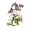
| ||||||||
| 2 | 
| ||||||||
| Unit cell |
|
-Components
| #1: Protein | / MMP-14 / Membrane-type matrix metalloproteinase 1 / MT-MMP 1 / MTMMP1 / Membrane-type-1 matrix ...MMP-14 / Membrane-type matrix metalloproteinase 1 / MT-MMP 1 / MTMMP1 / Membrane-type-1 matrix metalloproteinase / MT1-MMP / MT1MMP / MMP-X1 Mass: 20561.676 Da / Num. of mol.: 2 / Fragment: Residues 112-292 Source method: isolated from a genetically manipulated source Source: (gene. exp.) Homo sapiens (human) / Gene: MMP14 / Plasmid: pET3a / Production host:  Escherichia coli (E. coli) / Strain (production host): BL21 Escherichia coli (E. coli) / Strain (production host): BL21References: UniProt: P50281, membrane-type matrix metalloproteinase-1#2: Protein | Matrix metalloproteinase inhibitor / Tissue inhibitor of metalloproteinases 1 / TIMP-1 / Erythroid-potentiating activity / EPA / ...Tissue inhibitor of metalloproteinases 1 / TIMP-1 / Erythroid-potentiating activity / EPA / Fibroblast collagenase inhibitor / Collagenase inhibitorMass: 14126.209 Da / Num. of mol.: 2 / Fragment: Residues 24-148 / Mutation: V27A,P29V,T121L Source method: isolated from a genetically manipulated source Source: (gene. exp.) Homo sapiens (human) / Gene: TIMP1, CLGI, TIMP / Plasmid: pET3a / Production host: Escherichia coli (E. coli) / Strain (production host): BL21 / References: UniProt: P01033#3: Chemical | ChemComp-CA /   Mass: 40.078 Da / Num. of mol.: 4 / Source method: obtained synthetically / Formula: Ca Mass: 40.078 Da / Num. of mol.: 4 / Source method: obtained synthetically / Formula: Ca#4: Chemical | ChemComp-ZN /   Mass: 65.409 Da / Num. of mol.: 4 / Source method: obtained synthetically / Formula: Zn Mass: 65.409 Da / Num. of mol.: 4 / Source method: obtained synthetically / Formula: Zn#5: Water | ChemComp-HOH / | Water Mass: 18.015 Da / Num. of mol.: 145 / Source method: isolated from a natural source / Formula: H2O Mass: 18.015 Da / Num. of mol.: 145 / Source method: isolated from a natural source / Formula: H2O |
|---|
-Experimental details
-Experiment
| Experiment | Method: X-RAY DIFFRACTION / Number of used crystals: 1 |
|---|
- Sample preparation
| Crystal | Density Matthews: 2.3 Å3/Da / Density % sol: 46.42 % |
|---|---|
| Crystal grow | Temperature: 292 K / Method: vapor diffusion, hanging drop / pH: 5.5 Details: 100 mM Bis-Tris pH 5.5, 20% PEG 3350, VAPOR DIFFUSION, HANGING DROP, temperature 292K |
-Data collection
| Diffraction | Mean temperature: 100 K |
|---|---|
| Diffraction source | Source: ROTATING ANODE / Type: RIGAKU RU300 / Wavelength: 1.5418 Å |
| Detector | Type: RIGAKU RAXIS IV++ / Detector: IMAGE PLATE / Date: Jan 28, 2010 / Details: mirrors |
| Radiation | Protocol: SINGLE WAVELENGTH / Monochromatic (M) / Laue (L): M / Scattering type: x-ray |
| Radiation wavelength | Wavelength: 1.5418 Å / Relative weight: 1 |
| Reflection | Resolution: 2.05→50 Å / Num. obs: 39106 / % possible obs: 98.5 % / Observed criterion σ(F): 0 / Observed criterion σ(I): 0 / Redundancy: 7.2 % / Biso Wilson estimate: 24.36 Å2 / Rmerge(I) obs: 0.105 / Rsym value: 0.103 / Net I/σ(I): 19.5 |
| Reflection shell | Resolution: 2.05→2.09 Å / Redundancy: 5.7 % / Rmerge(I) obs: 0.27 / Mean I/σ(I) obs: 3.2 / Num. unique all: 1787 / Rsym value: 0.237 / % possible all: 90.9 |
- Processing
| Software |
| |||||||||||||||||||||||||||||||||||||||||||||||||||||||||||||||||
|---|---|---|---|---|---|---|---|---|---|---|---|---|---|---|---|---|---|---|---|---|---|---|---|---|---|---|---|---|---|---|---|---|---|---|---|---|---|---|---|---|---|---|---|---|---|---|---|---|---|---|---|---|---|---|---|---|---|---|---|---|---|---|---|---|---|---|
| Refinement | Method to determine structure: MOLECULAR REPLACEMENT Starting model: 1BQQ Resolution: 2.05→50 Å / Cor.coef. Fo:Fc: 0.946 / Cor.coef. Fo:Fc free: 0.915 / SU B: 4.507 / SU ML: 0.124 / Cross valid method: THROUGHOUT / ESU R: 0.209 / ESU R Free: 0.184 / Stereochemistry target values: MAXIMUM LIKELIHOOD / Details: HYDROGENS HAVE BEEN ADDED IN THE RIDING POSITIONS
| |||||||||||||||||||||||||||||||||||||||||||||||||||||||||||||||||
| Solvent computation | Ion probe radii: 0.8 Å / Shrinkage radii: 0.8 Å / VDW probe radii: 1.4 Å / Solvent model: MASK | |||||||||||||||||||||||||||||||||||||||||||||||||||||||||||||||||
| Displacement parameters | Biso mean: 24.357 Å2
| |||||||||||||||||||||||||||||||||||||||||||||||||||||||||||||||||
| Refinement step | Cycle: LAST / Resolution: 2.05→50 Å
| |||||||||||||||||||||||||||||||||||||||||||||||||||||||||||||||||
| Refine LS restraints |
| |||||||||||||||||||||||||||||||||||||||||||||||||||||||||||||||||
| LS refinement shell | Resolution: 2.05→2.1 Å / Total num. of bins used: 20
|
