 Movie
Movie Controller


+ Open data

- Basic information
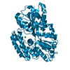
| Entry | Database: PDB / ID: 1bqq | ||||||
|---|---|---|---|---|---|---|---|
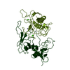
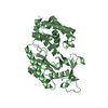
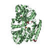
| Title | CRYSTAL STRUCTURE OF THE MT1-MMP--TIMP-2 COMPLEX | ||||||
 Components Components |
| ||||||
 Keywords Keywords | HYDROLASE/HYDROLASE INHIBITOR /  MATRIX METALLOPROTEINASE / TISSUE INHIBITOR OF METALLOPROTEINASES / PROTEINASE COMPLEX / PRO-GELATINASE A ACTIVATOR / COMPLEX (METALLOPROTEINASE-RECEPTOR) / HYDROLASE-HYDROLASE INHIBITOR COMPLEX MATRIX METALLOPROTEINASE / TISSUE INHIBITOR OF METALLOPROTEINASES / PROTEINASE COMPLEX / PRO-GELATINASE A ACTIVATOR / COMPLEX (METALLOPROTEINASE-RECEPTOR) / HYDROLASE-HYDROLASE INHIBITOR COMPLEX | ||||||
| Function / homology |  Function and homology informationmembrane-type matrix metalloproteinase-1 / craniofacial suture morphogenesis / positive regulation of macrophage migration / macropinosome / response to odorant / head development / negative regulation of membrane protein ectodomain proteolysis / chondrocyte proliferation / astrocyte cell migration / tissue remodeling ...membrane-type matrix metalloproteinase-1 / craniofacial suture morphogenesis / positive regulation of macrophage migration / macropinosome / response to odorant / head development / negative regulation of membrane protein ectodomain proteolysis / chondrocyte proliferation / astrocyte cell migration / tissue remodeling / metalloendopeptidase inhibitor activity / negative regulation of focal adhesion assembly / positive regulation of protein processing / endochondral ossification / embryonic cranial skeleton morphogenesis / endothelial cell proliferation / zymogen activation / intermediate filament cytoskeleton / positive regulation of B cell differentiation / branching morphogenesis of an epithelial tube / positive regulation of myotube differentiation / negative regulation of Notch signaling pathway / Activation of Matrix Metalloproteinases / endodermal cell differentiation / metalloaminopeptidase activity / Collagen degradation / collagen catabolic process / extracellular matrix disassembly / regulation of protein localization to plasma membrane / response to mechanical stimulus / ovarian follicle development / Degradation of the extracellular matrix / extracellular matrix organization / response to hormone / extracellular matrix / skeletal system development / response to cytokine / cell motility / lung development / response to organic cyclic compound / protein processing / metalloendopeptidase activity / Golgi lumen / response to estrogen / male gonad development / melanosome / integrin binding / cytoplasmic vesicle / positive regulation of cell growth / angiogenesis / endopeptidase activity / response to oxidative stress / protease binding / response to hypoxia / positive regulation of cell migration / serine-type endopeptidase activity / focal adhesion / proteolysis / extracellular space / zinc ion binding / metal ion binding / nucleus / plasma membrane / cytosol Function and homology informationmembrane-type matrix metalloproteinase-1 / craniofacial suture morphogenesis / positive regulation of macrophage migration / macropinosome / response to odorant / head development / negative regulation of membrane protein ectodomain proteolysis / chondrocyte proliferation / astrocyte cell migration / tissue remodeling ...membrane-type matrix metalloproteinase-1 / craniofacial suture morphogenesis / positive regulation of macrophage migration / macropinosome / response to odorant / head development / negative regulation of membrane protein ectodomain proteolysis / chondrocyte proliferation / astrocyte cell migration / tissue remodeling / metalloendopeptidase inhibitor activity / negative regulation of focal adhesion assembly / positive regulation of protein processing / endochondral ossification / embryonic cranial skeleton morphogenesis / endothelial cell proliferation / zymogen activation / intermediate filament cytoskeleton / positive regulation of B cell differentiation / branching morphogenesis of an epithelial tube / positive regulation of myotube differentiation / negative regulation of Notch signaling pathway / Activation of Matrix Metalloproteinases / endodermal cell differentiation / metalloaminopeptidase activity / Collagen degradation / collagen catabolic process / extracellular matrix disassembly / regulation of protein localization to plasma membrane / response to mechanical stimulus / ovarian follicle development / Degradation of the extracellular matrix / extracellular matrix organization / response to hormone / extracellular matrix / skeletal system development / response to cytokine / cell motility / lung development / response to organic cyclic compound / protein processing / metalloendopeptidase activity / Golgi lumen / response to estrogen / male gonad development / melanosome / integrin binding / cytoplasmic vesicle / positive regulation of cell growth / angiogenesis / endopeptidase activity / response to oxidative stress / protease binding / response to hypoxia / positive regulation of cell migration / serine-type endopeptidase activity / focal adhesion / proteolysis / extracellular space / zinc ion binding / metal ion binding / nucleus / plasma membrane / cytosolSimilarity search - Function | ||||||
| Biological species |  Homo sapiens (human) Homo sapiens (human) Bos taurus (cattle) Bos taurus (cattle) | ||||||
| Method | X-RAY DIFFRACTION / MOLECULAR REPLACEMENT / Resolution: 2.75 Å | ||||||
 Authors Authors | Fernandez-Catalan, C. / Bode, W. / Huber, R. / Turk, D. / Calvete, J.J. / Lichte, A. / Tschesche, H. / Maskos, K. | ||||||
 Citation Citation | Journal: EMBO J. / Year: 1998 Title: Crystal structure of the complex formed by the membrane type 1-matrix metalloproteinase with the tissue inhibitor of metalloproteinases-2, the soluble progelatinase A receptor. Authors: Fernandez-Catalan, C. / Bode, W. / Huber, R. / Turk, D. / Calvete, J.J. / Lichte, A. / Tschesche, H. / Maskos, K. | ||||||
| History |
|
- Structure visualization
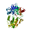
| Structure viewer | Molecule: MolmilJmol/JSmol |
|---|
- Downloads & links
-Download
| PDBx/mmCIF format | 1bqq.cif.gz | 89 KB | Display | PDBx/mmCIF format |
|---|---|---|---|---|
| PDB format | pdb1bqq.ent.gz | 71.2 KB | Display | PDB format |
| PDBx/mmJSON format | 1bqq.json.gz | Tree view | PDBx/mmJSON format | |
| Others |  Other downloads Other downloads |
-Validation report
| Arichive directory | https://data.pdbj.org/pub/pdb/validation_reports/bq/1bqqftp://data.pdbj.org/pub/pdb/validation_reports/bq/1bqq | HTTPS FTP |
|---|
-Related structure data


-Links
 PDBj
PDBj


- Assembly
| Deposited unit | 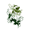
| ||||||||
|---|---|---|---|---|---|---|---|---|---|
| 1 |
| ||||||||
| Unit cell |
|
-Components
| #1: Protein | Mass: 19795.799 Da / Num. of mol.: 1 Source method: isolated from a genetically manipulated source Source: (gene. exp.) Homo sapiens (human) / Production host:  Escherichia coli (E. coli) Escherichia coli (E. coli)References: UniProt: P50281, Hydrolases; Acting on peptide bonds (peptidases); Metalloendopeptidases | ||||
|---|---|---|---|---|---|
| #2: Protein | Matrix metalloproteinase inhibitor / TIMP-2 / TISSUE INHIBITOR OF METALLOPROTEINASES-2 / COLLAGENASE INHIBITOR Mass: 20528.699 Da / Num. of mol.: 1 Source method: isolated from a genetically manipulated source Source: (gene. exp.) Bos taurus (cattle) / Production host: Escherichia coli (E. coli) / References: UniProt: P16368 | ||||
| #3: Chemical |   Mass: 40.078 Da / Num. of mol.: 2 / Source method: obtained synthetically / Formula: Ca Mass: 40.078 Da / Num. of mol.: 2 / Source method: obtained synthetically / Formula: Ca#4: Chemical |   Mass: 65.409 Da / Num. of mol.: 2 / Source method: obtained synthetically / Formula: Zn Mass: 65.409 Da / Num. of mol.: 2 / Source method: obtained synthetically / Formula: Zn#5: Water | ChemComp-HOH / | Water Mass: 18.015 Da / Num. of mol.: 311 / Source method: isolated from a natural source / Formula: H2O Mass: 18.015 Da / Num. of mol.: 311 / Source method: isolated from a natural source / Formula: H2O |
-Experimental details
-Experiment
| Experiment | Method: X-RAY DIFFRACTION / Number of used crystals: 1 |
|---|
- Sample preparation
| Crystal | Density Matthews: 2.03 Å3/Da / Density % sol: 40 % | |||||||||||||||||||||||||||||||||||||||||||||||||||||||||||||||
|---|---|---|---|---|---|---|---|---|---|---|---|---|---|---|---|---|---|---|---|---|---|---|---|---|---|---|---|---|---|---|---|---|---|---|---|---|---|---|---|---|---|---|---|---|---|---|---|---|---|---|---|---|---|---|---|---|---|---|---|---|---|---|---|---|
| Crystal grow | pH: 5.6 / Details: pH 5.60 | |||||||||||||||||||||||||||||||||||||||||||||||||||||||||||||||
| Crystal grow | *PLUS pH: 5.6 / Method: vapor diffusion | |||||||||||||||||||||||||||||||||||||||||||||||||||||||||||||||
| Components of the solutions | *PLUS
|
-Data collection
| Diffraction | Mean temperature: 300 K |
|---|---|
| Diffraction source | Source: ROTATING ANODE / Wavelength: 1.5418 |
| Detector | Type: MARRESEARCH / Detector: IMAGE PLATE / Date: Apr 15, 1997 |
| Radiation | Protocol: SINGLE WAVELENGTH / Monochromatic (M) / Laue (L): M / Scattering type: x-ray |
| Radiation wavelength | Wavelength: 1.5418 Å / Relative weight: 1 |
| Reflection | Resolution: 2.5→20 Å / Num. obs: 11911 / % possible obs: 97 % / Observed criterion σ(I): 2 / Redundancy: 2 % / Rmerge(I) obs: 0.095 |
| Reflection shell | Resolution: 2.5→2.6 Å / Rmerge(I) obs: 0.389 / % possible all: 93.2 |
| Reflection | *PLUS Highest resolution: 2.5 Å / Lowest resolution: 20 Å / Num. measured all: 73215 |
| Reflection shell | *PLUS % possible obs: 93.2 % |
- Processing
| Software |
| ||||||||||||||||||||||||||||||||||||||||||||||||||||||||||||
|---|---|---|---|---|---|---|---|---|---|---|---|---|---|---|---|---|---|---|---|---|---|---|---|---|---|---|---|---|---|---|---|---|---|---|---|---|---|---|---|---|---|---|---|---|---|---|---|---|---|---|---|---|---|---|---|---|---|---|---|---|---|
| Refinement | Method to determine structure: MOLECULAR REPLACEMENT / Resolution: 2.75→20 Å / Data cutoff high absF: 1000000 / Data cutoff low absF: 0.001 / Cross valid method: THROUGHOUT / σ(F): 2
| ||||||||||||||||||||||||||||||||||||||||||||||||||||||||||||
| Refinement step | Cycle: LAST / Resolution: 2.75→20 Å
| ||||||||||||||||||||||||||||||||||||||||||||||||||||||||||||
| Refine LS restraints |
| ||||||||||||||||||||||||||||||||||||||||||||||||||||||||||||
| Software | *PLUS Name: X-PLOR / Version: 3.843 / Classification: refinement | ||||||||||||||||||||||||||||||||||||||||||||||||||||||||||||
| Refinement | *PLUS Lowest resolution: 8 Å / Num. reflection obs: 8537 | ||||||||||||||||||||||||||||||||||||||||||||||||||||||||||||
| Solvent computation | *PLUS | ||||||||||||||||||||||||||||||||||||||||||||||||||||||||||||
| Displacement parameters | *PLUS |
