 Movie
Movie Controller

[English] 日本語
 Yorodumi
- PDB-3lto: Crystal structure of a mevalonate diphosphate decarboxylase from ... -
+ Open data

- Basic information
| Entry | Database: PDB / ID: 3lto | ||||||
|---|---|---|---|---|---|---|---|
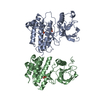
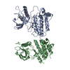
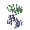
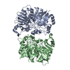
| Title | Crystal structure of a mevalonate diphosphate decarboxylase from Legionella pneumophila | ||||||
 Components Components | Mevalonate diphosphate decarboxylase | ||||||
 Keywords Keywords |  LYASE / Mevalonate diphosphate decarboxylase / Legionella pneumophila / Protein Structure Initiative II(PSI II) / NYSGXRC / 11277d / Structural Genomics / New York SGX Research Center for Structural Genomics / ATP-binding / Kinase / Nucleotide-binding / Transferase LYASE / Mevalonate diphosphate decarboxylase / Legionella pneumophila / Protein Structure Initiative II(PSI II) / NYSGXRC / 11277d / Structural Genomics / New York SGX Research Center for Structural Genomics / ATP-binding / Kinase / Nucleotide-binding / Transferase | ||||||
| Function / homology |  Function and homology informationdiphosphomevalonate decarboxylase / diphosphomevalonate decarboxylase activity / isoprenoid biosynthetic process / kinase activity Function and homology informationdiphosphomevalonate decarboxylase / diphosphomevalonate decarboxylase activity / isoprenoid biosynthetic process / kinase activitySimilarity search - Function | ||||||
| Biological species |   Legionella pneumophila (bacteria) Legionella pneumophila (bacteria) | ||||||
| Method | X-RAY DIFFRACTION / SYNCHROTRON / SAD / Resolution: 2.27 Å | ||||||
 Authors Authors | Palani, K. / Burley, S.K. / Swaminathan, S. / New York SGX Research Center for Structural Genomics (NYSGXRC) | ||||||
 Citation Citation | Journal: To be Published Title: Crystal structure of a mevalonate diphosphate decarboxylase from Legionella pneumophila Authors: Palani, K. / Burley, S.K. / Swaminathan, S. | ||||||
| History |
|
- Structure visualization
| Structure viewer | Molecule: MolmilJmol/JSmol |
|---|
- Downloads & links
-Download
| PDBx/mmCIF format | 3lto.cif.gz | 137 KB | Display | PDBx/mmCIF format |
|---|---|---|---|---|
| PDB format | pdb3lto.ent.gz | 111.9 KB | Display | PDB format |
| PDBx/mmJSON format | 3lto.json.gz | Tree view | PDBx/mmJSON format | |
| Others |  Other downloads Other downloads |
-Validation report
| Arichive directory | https://data.pdbj.org/pub/pdb/validation_reports/lt/3ltoftp://data.pdbj.org/pub/pdb/validation_reports/lt/3lto | HTTPS FTP |
|---|
-Related structure data
| Similar structure data | |
|---|---|
| Other databases |


-Links
 PDBj
PDBj- Assembly
| Deposited unit | 
| ||||||||
|---|---|---|---|---|---|---|---|---|---|
| 1 |
| ||||||||
| 2 | 
| ||||||||
| 3 | 
| ||||||||
| Unit cell |
|
-Components
| #1: Protein | Mass: 36793.984 Da / Num. of mol.: 2 Source method: isolated from a genetically manipulated source Source: (gene. exp.) Legionella pneumophila (bacteria) / Strain: Philadelphia 1/ATCC 33152/DSM 7513 / Gene: lpg2040 / Plasmid: BC-pSGX3(BC) / Production host: Escherichia coli (E. coli) / Strain (production host): BL21(DE3)References: UniProt: Q5ZTW8, diphosphomevalonate decarboxylase#2: Chemical | ChemComp-SO4 / Sulfate  Mass: 96.063 Da / Num. of mol.: 6 / Source method: obtained synthetically / Formula: SO4 Mass: 96.063 Da / Num. of mol.: 6 / Source method: obtained synthetically / Formula: SO4#3: Water | ChemComp-HOH / | Water Mass: 18.015 Da / Num. of mol.: 235 / Source method: isolated from a natural source / Formula: H2O Mass: 18.015 Da / Num. of mol.: 235 / Source method: isolated from a natural source / Formula: H2O |
|---|
-Experimental details
-Experiment
| Experiment | Method: X-RAY DIFFRACTION / Number of used crystals: 1 |
|---|
- Sample preparation
| Crystal | Density Matthews: 2.2 Å3/Da / Density % sol: 44.02 % |
|---|---|
| Crystal grow | Temperature: 298 K / Method: vapor diffusion, sitting drop / pH: 4.6 Details: 0.2M Ammonium Sulfate, 0.1M Sodium Acetate trihydrate, 30% PEG MME 2000, pH 4.6, VAPOR DIFFUSION, SITTING DROP, temperature 298.0K |
-Data collection
| Diffraction | Mean temperature: 100 K |
|---|---|
| Diffraction source | Source: SYNCHROTRON / Site: NSLS  / Beamline: X12C / Wavelength: 0.979 Å / Beamline: X12C / Wavelength: 0.979 Å |
| Detector | Type: ADSC QUANTUM 210 / Detector: CCD / Date: Nov 20, 2009 / Details: MIRRORS |
| Radiation | Monochromator: Si(III) Channel / Protocol: SINGLE WAVELENGTH / Monochromatic (M) / Laue (L): M / Scattering type: x-ray |
| Radiation wavelength | Wavelength: 0.979 Å / Relative weight: 1 |
| Reflection | Resolution: 2.27→40.9 Å / Num. all: 31363 / Num. obs: 30157 / % possible obs: 99.6 % / Observed criterion σ(F): 0 / Observed criterion σ(I): 0 / Redundancy: 14.5 % / Biso Wilson estimate: 28.5 Å2 / Rmerge(I) obs: 0.12 / Net I/σ(I): 7.6 |
| Reflection shell | Resolution: 2.27→2.35 Å / Redundancy: 13 % / Rmerge(I) obs: 0.393 / Mean I/σ(I) obs: 2.5 / Num. unique all: 2954 / % possible all: 96.1 |
- Processing
| Software |
| |||||||||||||||||||||||||
|---|---|---|---|---|---|---|---|---|---|---|---|---|---|---|---|---|---|---|---|---|---|---|---|---|---|---|
| Refinement | Method to determine structure: SAD / Resolution: 2.27→40.86 Å / Rfactor Rfree error: 0.007 / Data cutoff high absF: 39870.54 / Data cutoff low absF: 0 / Isotropic thermal model: RESTRAINED / Cross valid method: THROUGHOUT / σ(F): 0 / Stereochemistry target values: Engh & Huber
| |||||||||||||||||||||||||
| Solvent computation | Solvent model: FLAT MODEL / Bsol: 25.1317 Å2 / ksol: 0.366639 e/Å3 | |||||||||||||||||||||||||
| Displacement parameters | Biso mean: 18.3 Å2
| |||||||||||||||||||||||||
| Refine analyze |
| |||||||||||||||||||||||||
| Refinement step | Cycle: LAST / Resolution: 2.27→40.86 Å
| |||||||||||||||||||||||||
| Refine LS restraints |
| |||||||||||||||||||||||||
| Refine LS restraints NCS | NCS model details: NONE | |||||||||||||||||||||||||
| LS refinement shell | Resolution: 2.27→2.41 Å / Rfactor Rfree error: 0.019 / Total num. of bins used: 6
| |||||||||||||||||||||||||
| Xplor file |
|
