 Movie
Movie Controller

[English] 日本語
 Yorodumi
- PDB-3uws: Crystal structure of a clostripain (PARMER_00083) from Parabacter... -
+ Open data

- Basic information
| Entry | Database: PDB / ID: 3uws | ||||||
|---|---|---|---|---|---|---|---|
| Title | Crystal structure of a clostripain (PARMER_00083) from Parabacteroides merdae ATCC 43184 at 1.70 A resolution | ||||||
 Components Components | (hypothetical protein ) x 2 ) x 2 | ||||||
 Keywords Keywords | Structural Genomics / Unknown Function / clostripain family protein / Peptidase_C11 / Joint Center for Structural Genomics / JCSG / Protein Structure Initiative / PSI-BIOLOGY | ||||||
| Function / homology | Rossmann fold - #11970 / Peptidase C11, clostripain / Clostripain family / Prokaryotic membrane lipoprotein lipid attachment site profile. / Rossmann fold / 3-Layer(aba) Sandwich / Alpha Beta / Clostripain family Function and homology information Function and homology information | ||||||
| Biological species |  Parabacteroides merdae (bacteria) Parabacteroides merdae (bacteria) | ||||||
| Method | X-RAY DIFFRACTION / SYNCHROTRON / SAD / Resolution: 1.7 Å | ||||||
 Authors Authors | Joint Center for Structural Genomics (JCSG) | ||||||
 Citation Citation | Journal: J.Biol.Chem. / Year: 2016 Title: Crystal Structure and Activity Studies of the C11 Cysteine Peptidase from Parabacteroides merdae in the Human Gut Microbiome. Authors: McLuskey, K. / Grewal, J.S. / Das, D. / Godzik, A. / Lesley, S.A. / Deacon, A.M. / Coombs, G.H. / Elsliger, M.A. / Wilson, I.A. / Mottram, J.C. | ||||||
| History |
|
- Structure visualization
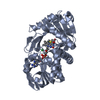
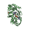
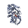
| Structure viewer | Molecule: MolmilJmol/JSmol |
|---|
- Downloads & links
-Download
| PDBx/mmCIF format | 3uws.cif.gz | 307.7 KB | Display | PDBx/mmCIF format |
|---|---|---|---|---|
| PDB format | pdb3uws.ent.gz | 254.2 KB | Display | PDB format |
| PDBx/mmJSON format | 3uws.json.gz | Tree view | PDBx/mmJSON format | |
| Others |  Other downloads Other downloads |
-Validation report
| Arichive directory | https://data.pdbj.org/pub/pdb/validation_reports/uw/3uwsftp://data.pdbj.org/pub/pdb/validation_reports/uw/3uws | HTTPS FTP |
|---|
-Related structure data
| Similar structure data | |
|---|---|
| Other databases |


-Links
 PDBj
PDBj


- Assembly
| Deposited unit | 
| ||||||||
|---|---|---|---|---|---|---|---|---|---|
| 1 | 
| ||||||||
| 2 | 
| ||||||||
| Unit cell |
|
-Components
| #1: Protein | Mass: 14162.332 Da / Num. of mol.: 2 / Fragment: UNP residues 23-147 Source method: isolated from a genetically manipulated source Source: (gene. exp.) Parabacteroides merdae (bacteria) / Strain: ATCC 43184 / Gene: PARMER_00083 / Plasmid: SpeedET / Production host: Escherichia coli (E. coli) / Strain (production host): PB1 / References: UniProt: A7A9N3#2: Protein | Mass: 26838.033 Da / Num. of mol.: 2 / Fragment: UNP residues 148-375 Source method: isolated from a genetically manipulated source Source: (gene. exp.) Parabacteroides merdae (bacteria) / Strain: ATCC 43184 / Gene: PARMER_00083 / Plasmid: SpeedET / Production host: Escherichia coli (E. coli) / Strain (production host): PB1 / References: UniProt: A7A9N3#3: Chemical | ChemComp-EDO / Ethylene glycol  Mass: 62.068 Da / Num. of mol.: 7 / Source method: obtained synthetically / Formula: C2H6O2 Mass: 62.068 Da / Num. of mol.: 7 / Source method: obtained synthetically / Formula: C2H6O2#4: Water | ChemComp-HOH / | Water Mass: 18.015 Da / Num. of mol.: 690 / Source method: isolated from a natural source / Formula: H2O Mass: 18.015 Da / Num. of mol.: 690 / Source method: isolated from a natural source / Formula: H2OCompound details | THERE IS A BREAK IN THE CHAIN BETWEEN RESIDUES 147 AND 148. CLEAVAGE AT THIS SITE WAS CONFIRMED BY ...THERE IS A BREAK IN THE CHAIN BETWEEN RESIDUES 147 AND 148. CLEAVAGE AT THIS SITE WAS CONFIRMED BY INTACT MASS SPECTROMET | Sequence details | THIS CONSTRUCT (RESIDUES 23-375) WAS EXPRESSED WITH A PURIFICATION TAG MGSDKIHHHHHHENLYFQG. THE TAG ...THIS CONSTRUCT (RESIDUES 23-375) WAS EXPRESSED WITH A PURIFICATI | |
|---|
-Experimental details
-Experiment
| Experiment | Method: X-RAY DIFFRACTION / Number of used crystals: 1 |
|---|
- Sample preparation
| Crystal | Density Matthews: 2.02 Å3/Da / Density % sol: 38.96 % |
|---|---|
| Crystal grow | Temperature: 277 K / Method: vapor diffusion, sitting drop / pH: 6.3 Details: 0.2M NH4Cl, 20.0% PEG-3350, No Buffer pH 6.3, NANODROP, VAPOR DIFFUSION, SITTING DROP, temperature 277K |
-Data collection
| Diffraction | Mean temperature: 100 K | |||||||||||||||||||||||||||||||||||||||||||||||||||||||||||||||||||||||||||||
|---|---|---|---|---|---|---|---|---|---|---|---|---|---|---|---|---|---|---|---|---|---|---|---|---|---|---|---|---|---|---|---|---|---|---|---|---|---|---|---|---|---|---|---|---|---|---|---|---|---|---|---|---|---|---|---|---|---|---|---|---|---|---|---|---|---|---|---|---|---|---|---|---|---|---|---|---|---|---|
| Diffraction source | Source: SYNCHROTRON / Site: ALS  / Beamline: 8.2.2 / Wavelength: 0.9793 / Beamline: 8.2.2 / Wavelength: 0.9793 | |||||||||||||||||||||||||||||||||||||||||||||||||||||||||||||||||||||||||||||
| Detector | Type: ADSC QUANTUM 315 / Detector: CCD / Date: Oct 15, 2011 / Details: KOHZU: Double Crystal Si(111) | |||||||||||||||||||||||||||||||||||||||||||||||||||||||||||||||||||||||||||||
| Radiation | Monochromator: Double Crystal Si(111) / Protocol: SINGLE WAVELENGTH / Monochromatic (M) / Laue (L): M / Scattering type: x-ray | |||||||||||||||||||||||||||||||||||||||||||||||||||||||||||||||||||||||||||||
| Radiation wavelength | Wavelength: 0.9793 Å / Relative weight: 1 | |||||||||||||||||||||||||||||||||||||||||||||||||||||||||||||||||||||||||||||
| Reflection | Resolution: 1.7→28.734 Å / Num. obs: 70913 / % possible obs: 99.1 % / Observed criterion σ(I): -3 / Biso Wilson estimate: 15.869 Å2 / Rmerge(I) obs: 0.08 / Net I/σ(I): 11.34 | |||||||||||||||||||||||||||||||||||||||||||||||||||||||||||||||||||||||||||||
| Reflection shell | Diffraction-ID: 1
|
-Phasing
| Phasing | Method: SAD |
|---|
- Processing
| Software |
| |||||||||||||||||||||||||||||||||||||||||||||||||||||||||||||||||||||||||||||||||||||||||||||||||||||||||||||||||||||||||||||
|---|---|---|---|---|---|---|---|---|---|---|---|---|---|---|---|---|---|---|---|---|---|---|---|---|---|---|---|---|---|---|---|---|---|---|---|---|---|---|---|---|---|---|---|---|---|---|---|---|---|---|---|---|---|---|---|---|---|---|---|---|---|---|---|---|---|---|---|---|---|---|---|---|---|---|---|---|---|---|---|---|---|---|---|---|---|---|---|---|---|---|---|---|---|---|---|---|---|---|---|---|---|---|---|---|---|---|---|---|---|---|---|---|---|---|---|---|---|---|---|---|---|---|---|---|---|---|
| Refinement | Method to determine structure: SAD / Resolution: 1.7→28.734 Å / Cor.coef. Fo:Fc: 0.971 / Cor.coef. Fo:Fc free: 0.96 / Occupancy max: 1 / Occupancy min: 0.25 / SU B: 3.587 / SU ML: 0.06 / Cross valid method: THROUGHOUT / σ(F): 0 / ESU R: 0.098 / ESU R Free: 0.095 Stereochemistry target values: MAXIMUM LIKELIHOOD WITH PHASES Details: 1.HYDROGENS HAVE BEEN ADDED IN THE RIDING POSITIONS. 2.A MET-INHIBITION PROTOCOL WAS USED FOR SELENOMETHIONINE INCORPORATION DURING PROTEIN EXPRESSION. THE OCCUPANCY OF THE SE ATOMS IN THE ...Details: 1.HYDROGENS HAVE BEEN ADDED IN THE RIDING POSITIONS. 2.A MET-INHIBITION PROTOCOL WAS USED FOR SELENOMETHIONINE INCORPORATION DURING PROTEIN EXPRESSION. THE OCCUPANCY OF THE SE ATOMS IN THE MSE RESIDUES WAS REDUCED TO 0.75 FOR THE REDUCED SCATTERING POWER DUE TO PARTIAL S-MET INCORPORATION. 3.ATOM RECORD CONTAINS SUM OF TLS AND RESIDUAL B FACTORS. ANISOU RECORD CONTAINS SUM OF TLS AND RESIDUAL U FACTORS. 4.WATERS WERE EXCLUDED FROM AUTOMATIC TLS ASSIGNMENT. 5.1,2-ETHANEDIOL (EDO) FROM THE CRYOPROTECTANT SOLUTION HAS BEEN MODELED IN THE SOLVENT STRUCTURE. 6.THE PROTEIN IS CLEAVED AFTER RESIDUE 147, WHICH RESULTS IN RESIDUE 148 BEING LOCATED FAR FROM RESIDUE 147.
| |||||||||||||||||||||||||||||||||||||||||||||||||||||||||||||||||||||||||||||||||||||||||||||||||||||||||||||||||||||||||||||
| Solvent computation | Ion probe radii: 0.8 Å / Shrinkage radii: 0.8 Å / VDW probe radii: 1.2 Å / Solvent model: BABINET MODEL WITH MASK | |||||||||||||||||||||||||||||||||||||||||||||||||||||||||||||||||||||||||||||||||||||||||||||||||||||||||||||||||||||||||||||
| Displacement parameters | Biso max: 65.01 Å2 / Biso mean: 19.9673 Å2 / Biso min: 7.45 Å2
| |||||||||||||||||||||||||||||||||||||||||||||||||||||||||||||||||||||||||||||||||||||||||||||||||||||||||||||||||||||||||||||
| Refinement step | Cycle: LAST / Resolution: 1.7→28.734 Å
| |||||||||||||||||||||||||||||||||||||||||||||||||||||||||||||||||||||||||||||||||||||||||||||||||||||||||||||||||||||||||||||
| Refine LS restraints |
| |||||||||||||||||||||||||||||||||||||||||||||||||||||||||||||||||||||||||||||||||||||||||||||||||||||||||||||||||||||||||||||
| LS refinement shell | Resolution: 1.7→1.744 Å / Total num. of bins used: 20
| |||||||||||||||||||||||||||||||||||||||||||||||||||||||||||||||||||||||||||||||||||||||||||||||||||||||||||||||||||||||||||||
| Refinement TLS params. | Method: refined / Refine-ID: X-RAY DIFFRACTION
| |||||||||||||||||||||||||||||||||||||||||||||||||||||||||||||||||||||||||||||||||||||||||||||||||||||||||||||||||||||||||||||
| Refinement TLS group |
|
