 Movie
Movie Controller


+ Open data

- Basic information
| Entry | Database: PDB / ID: 4myg | ||||||
|---|---|---|---|---|---|---|---|
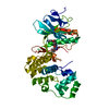
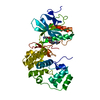
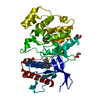
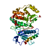
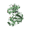
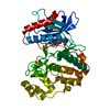
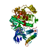
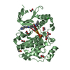
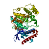
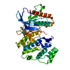
| Title | MAPK13, active form | ||||||
 Components Components | Mitogen-activated protein kinase 13 P38 mitogen-activated protein kinases P38 mitogen-activated protein kinases | ||||||
 Keywords Keywords | TRANSFERASE / p38 kinase / phosphorylation | ||||||
| Function / homology |  Function and homology information Function and homology informationcellular response to anisomycin / cellular response to sorbitol / cellular response to sodium arsenite / response to osmotic stress / DSCAM interactions / MAP kinase activity / mitogen-activated protein kinase / cellular response to interleukin-1 / stress-activated MAPK cascade / p38MAPK events ...cellular response to anisomycin / cellular response to sorbitol / cellular response to sodium arsenite / response to osmotic stress / DSCAM interactions / MAP kinase activity / mitogen-activated protein kinase / cellular response to interleukin-1 / stress-activated MAPK cascade / p38MAPK events / NOD1/2 Signaling Pathway / VEGFA-VEGFR2 Pathway / cellular response to hydrogen peroxide / positive regulation of inflammatory response / positive regulation of interleukin-6 production / cellular response to UV / peptidyl-serine phosphorylation / intracellular signal transduction / cell cycle / protein serine kinase activity / protein serine/threonine kinase activity / ATP binding / nucleus / cytosol / cytoplasmSimilarity search - Function | ||||||
| Biological species |  Homo sapiens (human) Homo sapiens (human) | ||||||
| Method | X-RAY DIFFRACTION / SYNCHROTRON / MOLECULAR REPLACEMENT / Resolution: 2.594 Å | ||||||
 Authors Authors | Yurtsever, Z. / Brett, T.J. / Scheaffer, S.M. | ||||||
 Citation Citation | Journal: Acta Crystallogr.,Sect.D / Year: 2015 Title: The crystal structure of phosphorylated MAPK13 reveals common structural features and differences in p38 MAPK family activation. Authors: Yurtsever, Z. / Scheaffer, S.M. / Romero, A.G. / Holtzman, M.J. / Brett, T.J. | ||||||
| History |
|
- Structure visualization
| Structure viewer | Molecule: MolmilJmol/JSmol |
|---|
- Downloads & links
-Download
| PDBx/mmCIF format | 4myg.cif.gz | 152.3 KB | Display | PDBx/mmCIF format |
|---|---|---|---|---|
| PDB format | pdb4myg.ent.gz | 119.7 KB | Display | PDB format |
| PDBx/mmJSON format | 4myg.json.gz | Tree view | PDBx/mmJSON format | |
| Others |  Other downloads Other downloads |
-Validation report
| Arichive directory | https://data.pdbj.org/pub/pdb/validation_reports/my/4mygftp://data.pdbj.org/pub/pdb/validation_reports/my/4myg | HTTPS FTP |
|---|
-Related structure data
| Related structure data |  1cm8S  4mox 4mp5 4mp9 S: Starting model for refinement |
|---|---|
| Similar structure data |


-Links
 PDBj
PDBj


- Assembly
| Deposited unit | 
| ||||||||
|---|---|---|---|---|---|---|---|---|---|
| 1 | 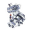
| ||||||||
| 2 | 
| ||||||||
| Unit cell |
|
-Components
| #1: Protein | P38 mitogen-activated protein kinases / MAP kinase 13 / MAPK 13 / Mitogen-activated protein kinase p38 delta / MAP kinase p38 delta / ...MAP kinase 13 / MAPK 13 / Mitogen-activated protein kinase p38 delta / MAP kinase p38 delta / Stress-activated protein kinase 4 Mass: 42932.004 Da / Num. of mol.: 2 / Fragment: UNP residues 1-352 Source method: isolated from a genetically manipulated source Source: (gene. exp.) Homo sapiens (human) / Gene: MAPK13, PRKM13, SAPK4 / Plasmid: pet28a / Production host:  Escherichia coli (E. coli) / Strain (production host): Rosetta2(DE3) Escherichia coli (E. coli) / Strain (production host): Rosetta2(DE3)References: UniProt: O15264, mitogen-activated protein kinase#2: Water | ChemComp-HOH / | Water Mass: 18.015 Da / Num. of mol.: 184 / Source method: isolated from a natural source / Formula: H2O Mass: 18.015 Da / Num. of mol.: 184 / Source method: isolated from a natural source / Formula: H2O |
|---|
-Experimental details
-Experiment
| Experiment | Method: X-RAY DIFFRACTION / Number of used crystals: 1 |
|---|
- Sample preparation
| Crystal | Density Matthews: 2.43 Å3/Da / Density % sol: 49.31 % |
|---|---|
| Crystal grow | Temperature: 290 K / Method: vapor diffusion, hanging drop / pH: 6.2 Details: 100 mM Bis-Tris, 21% PEG3350, 200 mM sodium chloride, pH 6.2, VAPOR DIFFUSION, HANGING DROP, temperature 290K |
-Data collection
| Diffraction | Mean temperature: 105 K |
|---|---|
| Diffraction source | Source: SYNCHROTRON / Site: ALS  / Beamline: 4.2.2 / Wavelength: 1.0007 Å / Beamline: 4.2.2 / Wavelength: 1.0007 Å |
| Detector | Type: NOIR-1 / Detector: CCD / Date: Aug 24, 2010 |
| Radiation | Monochromator: sagitally focused double crystal Si(111) / Protocol: SINGLE WAVELENGTH / Monochromatic (M) / Laue (L): M / Scattering type: x-ray |
| Radiation wavelength | Wavelength: 1.0007 Å / Relative weight: 1 |
| Reflection | Resolution: 2.594→50 Å / Num. all: 24975 / Num. obs: 24975 / % possible obs: 98.1 % / Observed criterion σ(F): 0 / Observed criterion σ(I): 0 / Redundancy: 8.9 % / Biso Wilson estimate: 48.55 Å2 / Rmerge(I) obs: 0.088 / Rsym value: 0.088 / Net I/σ(I): 19.8 |
| Reflection shell | Resolution: 2.594→2.69 Å / Redundancy: 5.5 % / Rmerge(I) obs: 0.59 / Mean I/σ(I) obs: 1.96 / Rsym value: 0.59 / % possible all: 90.3 |
- Processing
| Software |
| ||||||||||||||||||||||||||||||||||||||||||||||||||||||||||||||||||||||
|---|---|---|---|---|---|---|---|---|---|---|---|---|---|---|---|---|---|---|---|---|---|---|---|---|---|---|---|---|---|---|---|---|---|---|---|---|---|---|---|---|---|---|---|---|---|---|---|---|---|---|---|---|---|---|---|---|---|---|---|---|---|---|---|---|---|---|---|---|---|---|---|
| Refinement | Method to determine structure: MOLECULAR REPLACEMENT Starting model: PDB ENTRY 1CM8 Resolution: 2.594→49.307 Å / SU ML: 0.35 / σ(F): 1.34 / Phase error: 27.29 / Stereochemistry target values: ML
| ||||||||||||||||||||||||||||||||||||||||||||||||||||||||||||||||||||||
| Solvent computation | Shrinkage radii: 0.9 Å / VDW probe radii: 1.11 Å / Solvent model: FLAT BULK SOLVENT MODEL | ||||||||||||||||||||||||||||||||||||||||||||||||||||||||||||||||||||||
| Refinement step | Cycle: LAST / Resolution: 2.594→49.307 Å
| ||||||||||||||||||||||||||||||||||||||||||||||||||||||||||||||||||||||
| Refine LS restraints |
| ||||||||||||||||||||||||||||||||||||||||||||||||||||||||||||||||||||||
| LS refinement shell |
|
