 Movie
Movie Controller


+ Open data

- Basic information
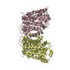
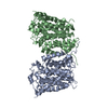
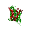
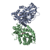
| Entry | Database: PDB / ID: 3lrb | |||||||||
|---|---|---|---|---|---|---|---|---|---|---|
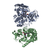
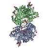
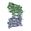
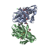
| Title | Structure of E. coli AdiC | |||||||||
 Components Components | Arginine/agmatine antiporter | |||||||||
 Keywords Keywords |  TRANSPORT PROTEIN / transporter / antiporter / AdiC / Amino-acid transport / Antiport / Cell inner membrane / Cell membrane / Membrane / Transmembrane / Transport TRANSPORT PROTEIN / transporter / antiporter / AdiC / Amino-acid transport / Antiport / Cell inner membrane / Cell membrane / Membrane / Transmembrane / Transport | |||||||||
| Function / homology | Amino acid/polyamine transporter I / Amino acid permease / amino acid transport / antiporter activity / identical protein binding / plasma membrane / Arginine/agmatine antiporter Function and homology information Function and homology information | |||||||||
| Biological species |  Escherichia coli (E. coli) Escherichia coli (E. coli) | |||||||||
| Method | X-RAY DIFFRACTION / SYNCHROTRON / SAD / Resolution: 3.61 Å | |||||||||
 Authors Authors | Gao, X. / Lu, F. / Zhou, L. / Shi, Y. | |||||||||
 Citation Citation | Journal: Science / Year: 2009 Title: Structure and mechanism of an amino acid antiporter Authors: Gao, X. / Lu, F. / Zhou, L. / Dang, S. / Sun, L. / Li, X. / Wang, J. / Shi, Y. | |||||||||
| History |
|
- Structure visualization
| Structure viewer | Molecule: MolmilJmol/JSmol |
|---|
- Downloads & links
-Download
| PDBx/mmCIF format | 3lrb.cif.gz | 321.1 KB | Display | PDBx/mmCIF format |
|---|---|---|---|---|
| PDB format | pdb3lrb.ent.gz | 267.3 KB | Display | PDB format |
| PDBx/mmJSON format | 3lrb.json.gz | Tree view | PDBx/mmJSON format | |
| Others |  Other downloads Other downloads |
-Validation report
| Arichive directory | https://data.pdbj.org/pub/pdb/validation_reports/lr/3lrbftp://data.pdbj.org/pub/pdb/validation_reports/lr/3lrb | HTTPS FTP |
|---|
-Related structure data


-Links
 PDBj
PDBj- Assembly
| Deposited unit | 
| ||||||||||||||||||||||||||||||||||||||||||||||||||||||||||||||||
|---|---|---|---|---|---|---|---|---|---|---|---|---|---|---|---|---|---|---|---|---|---|---|---|---|---|---|---|---|---|---|---|---|---|---|---|---|---|---|---|---|---|---|---|---|---|---|---|---|---|---|---|---|---|---|---|---|---|---|---|---|---|---|---|---|---|
| 1 |
| ||||||||||||||||||||||||||||||||||||||||||||||||||||||||||||||||
| Unit cell |
| ||||||||||||||||||||||||||||||||||||||||||||||||||||||||||||||||
| Noncrystallographic symmetry (NCS) | NCS domain:
NCS domain segments: Ens-ID: 1
|
-Components
| #1: Protein | Mass: 46869.258 Da / Num. of mol.: 2 Source method: isolated from a genetically manipulated source Source: (gene. exp.) Escherichia coli (E. coli) / Strain: O157:H7 / Gene: adiC, Z5717, ECs5097 / Plasmid: pET15b / Production host: Escherichia coli (E. coli) / Strain (production host): BL21(DE3) / References: UniProt: P60063 |
|---|
-Experimental details
-Experiment
| Experiment | Method: X-RAY DIFFRACTION / Number of used crystals: 1 |
|---|
- Sample preparation
| Crystal | Density Matthews: 3.7 Å3/Da / Density % sol: 66.78 % |
|---|---|
| Crystal grow | Temperature: 291 K / Method: vapor diffusion, hanging drop / pH: 7 Details: 0.4% NG, 0.1mM Tris, 22% (w/v) PEG 400, pH 7.0, VAPOR DIFFUSION, HANGING DROP, temperature 291K , temperature 291.0K |
-Data collection
| Diffraction | Mean temperature: 100 K |
|---|---|
| Diffraction source | Source: SYNCHROTRON / Site: SPring-8  / Beamline: BL41XU / Wavelength: 1 Å / Beamline: BL41XU / Wavelength: 1 Å |
| Detector | Type: MARMOSAIC 225 mm CCD / Detector: CCD / Date: Jul 14, 2008 |
| Radiation | Protocol: SINGLE WAVELENGTH / Monochromatic (M) / Laue (L): M / Scattering type: x-ray |
| Radiation wavelength | Wavelength: 1 Å / Relative weight: 1 |
| Reflection | Resolution: 3.61→50 Å / Num. obs: 16598 / Redundancy: 10.8 % / Biso Wilson estimate: 152.36 Å2 / Rmerge(I) obs: 0.083 |
| Reflection shell | Resolution: 3.61→3.75 Å / Redundancy: 5.3 % / Rmerge(I) obs: 0.463 / % possible all: 99.9 |
- Processing
| Software |
| |||||||||||||||||||||||||||||||||||||||||||||||||||||||||||||||||||||||||||
|---|---|---|---|---|---|---|---|---|---|---|---|---|---|---|---|---|---|---|---|---|---|---|---|---|---|---|---|---|---|---|---|---|---|---|---|---|---|---|---|---|---|---|---|---|---|---|---|---|---|---|---|---|---|---|---|---|---|---|---|---|---|---|---|---|---|---|---|---|---|---|---|---|---|---|---|---|
| Refinement | Method to determine structure: SAD / Resolution: 3.61→49.323 Å / SU ML: 0.58 / Isotropic thermal model: Isotropic / σ(F): 1.33 / Phase error: 36.94 / Stereochemistry target values: MLHL
| |||||||||||||||||||||||||||||||||||||||||||||||||||||||||||||||||||||||||||
| Solvent computation | Shrinkage radii: 0.9 Å / VDW probe radii: 1.11 Å / Solvent model: FLAT BULK SOLVENT MODEL / Bsol: 150 Å2 / ksol: 0.273 e/Å3 | |||||||||||||||||||||||||||||||||||||||||||||||||||||||||||||||||||||||||||
| Displacement parameters | Biso mean: 188.562 Å2
| |||||||||||||||||||||||||||||||||||||||||||||||||||||||||||||||||||||||||||
| Refinement step | Cycle: LAST / Resolution: 3.61→49.323 Å
| |||||||||||||||||||||||||||||||||||||||||||||||||||||||||||||||||||||||||||
| Refine LS restraints |
| |||||||||||||||||||||||||||||||||||||||||||||||||||||||||||||||||||||||||||
| Refine LS restraints NCS |
| |||||||||||||||||||||||||||||||||||||||||||||||||||||||||||||||||||||||||||
| LS refinement shell |
| |||||||||||||||||||||||||||||||||||||||||||||||||||||||||||||||||||||||||||
| Refinement TLS params. | Method: refined / Refine-ID: X-RAY DIFFRACTION
| |||||||||||||||||||||||||||||||||||||||||||||||||||||||||||||||||||||||||||
| Refinement TLS group |
|
